Types of Bone Cells
The bones are a core component of a living body that holds the structure of muscles and organs. Learn the three types of bone cells, their history, structure, and functions.
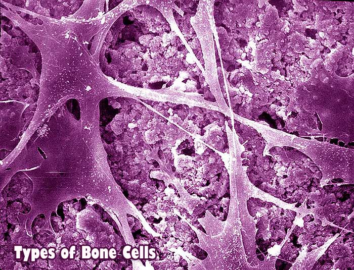
Types of Bone Cells: The bones are a core founding component of a living body that holds the structure of muscles and organs. The bones of the skeletal system is composed of two types of tissues, i.e., compact and spongy bone tissue.
The Compact bone tissue covers the outer part of the bone structure and provides toughness and strength to the structure of bone.
Spongy bone tissue is present inside the compact bone tissue. It is pretty soft and helps with compression that might occur as a result to stress on the bone.
Jump to:
Types of Bone Cells
The compact and spongy bone tissues are composed of 3 main types of bone cells. These bone cells have distinct features and structures and are considered essential functions. These bone cells are Osteoclasts, Osteoblasts, and Osteocytes.
These bone cells are embedded in the matrix of bony tissue and perform many vital functions.
From hormone production to the provision of mechanical strength, bone cells possess miraculous properties and functions fundamental to normal bone functioning.
This article gives an overview of the bone cells’ fundamental properties and some essential functions.
Osteoclasts
History of Osteoclasts
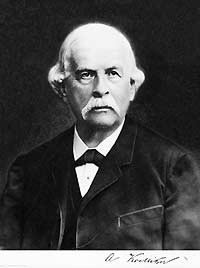
Osteoclasts are distinct bone cells doing bone demolishing work. They were discovered in 1873 by Albert von Kolliker.
- The resorption properties of osteoclasts inside the bony tissue were established in the early days of their discovery.
- During recent years, usage of the electron microscope and other technological advancements have helped in comprehending the behavior of osteoclasts in both healthy and diseased conditions.
- Osteoclasts consist of diverse ultrastructure and multiple nuclei. These features led the discoverers to study osteoclasts extensively under light and electron microscopes.
- Osteoclasts showed completely different behavior compared to other bone cells.
![]()
Structure of Osteoclasts
The occurrence of osteoclasts is quite scarce in the bony tissue.
- It is estimated that in an area of 1mm of the bony tissue, almost 2 to 3 osteoclasts are found.
- The structure of osteoclasts is related to their function. As osteoclasts have the fate of absorbing bone, they are giant bone cells with specialized membrane structures, ruffled borders, and clear zones, helping them function in bone resorption.
- Osteoclasts originate from liver and spleen as hematopoietic stem cells.
- After their release, osteoclast travels through the bloodstream to reach out to cells at the place of resorption.
At the time of their arrival at the resorption site, osteoclasts are composed of just one nucleus.
At the time of their early life, osteoclasts are closely related to other cells regarding the origin, such as macrophages, monocytes, and granulocytes.
These all cells are components of granulocyte-macrophage colony-forming a unit (GM-CFU).
Numerous research studies have been conducted to understand osteoclasts’ structure and their relationship with other closely related cells.
![]()
Function of Osteoclasts
The functions played by osteoclasts are crucial to the bone’s normal functioning.
- Osteoclasts regulate the homeostasis of the bone.
- If the number of osteoclasts gets lowered inside the bony tissue or they are not adequately developed, the bone dysfunctioning called Osteopetrosis develops.
- On the other hand, when osteoclasts are present in increased numbers than required, the bone problem of Osteoporosis may develop. Therefore, the number and amount of osteoclasts in the bone controls the bone structural integrity .
- Osteoclast resorbs bones by creating sealed compartments adjacent to the bone surface. Then, osteoclasts secrete acid phosphatases. These enzymes are acidic and function to degrade the bone. The resulting edge and depression on the bone formed by the action of these enzymes are termed as “ruffled border“.
- At the site of ruffled borders, osteoclasts are acted upon by carbonic anhydrase enzyme, and hydrogen ions are released as a result of a chemical reaction between CO2 and H2O.
- The resultant products of the reaction create an acidic environment at the site of ruffled borders and help in dissolving the bone, as shown in the equation: CO2 + H2O → H+ + HCO3-. The dissolved bone is converted into a matrix of calcium.
![]()
Osteoblasts
History of Osteoblasts

Osteoblasts are bone cells with a relatively different structure than other bone cells.
- They are short-lived cells. In the late 1950s, Harold M.Frost was involved in extensively studying the properties and behavior of osteoblasts.
- Frost discovered the collaborative nature of osteoblasts to work with osteoclasts in forming the bone matrix.
- He added through various experimentations that osteoblasts strengthen the integrity of the bony tissue.
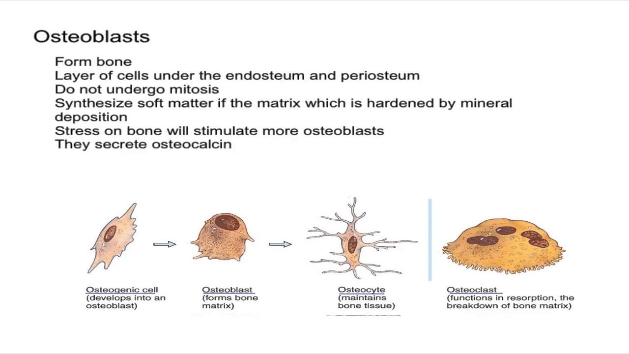
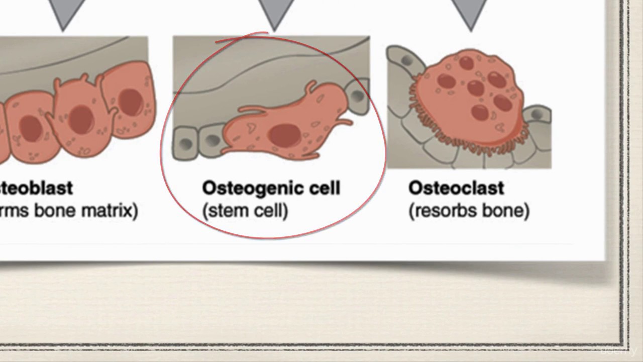
- Osteoblasts are also derived from bone marrow precursor cells (mesenchymal stem cells MSC). Osteogenic cells are undifferentiated and develop into Osteoblasts.
![]()
Structure of Osteoblasts
Osteoblasts are the bone cells possessing cuboidal and columnar shapes.
- They possess a central nucleus found on the bone’s surface, i.e., present in the old compact part of the bone.
- It is reported that osteoblast compromise about 6% of total bone cells.
- Osteoblasts are made from mesenchymal stem cells (MSC) along with muscle cells (myocytes) and fat cells (adipocytes).
- Osteoblasts work together in clusters and perform their function of building up the bone.
- Once the cluster of osteoblasts finishes its work, the shape of osteoblasts gets flattened.
- They are referred to as inactive osteoblasts at this stage. When osteoblasts become inactive, they transport toward the outer surface of the bone and settle along the line.
- This phenomenon gives osteoblasts a specific name for lining cells.
Following image represents the histological view of osteoblast after staining:
Osteoblast bone cells have some morphological features that closely resemble protein-synthesizing cells.
Protein synthesizing cells are characterized by the presence of prominent Golgi complex, rough endoplasmic reticulum along with many resembling secretory vesicles.
![]()
Function of Osteoblasts
Osteoblasts majorly perform two varieties of functions within the bone tissue.
Firstly, osteoblasts release multiple proteins essential to the formation of the bony structure matrix.
Secondly, osteoblasts help in regulating the mineralization of bone. Some of the primary functions of osteoblasts are mentioned below:
- Osteoblasts have special receptors for hormones such as estrogen, parathyroid hormone (PTH), and vitamin D.
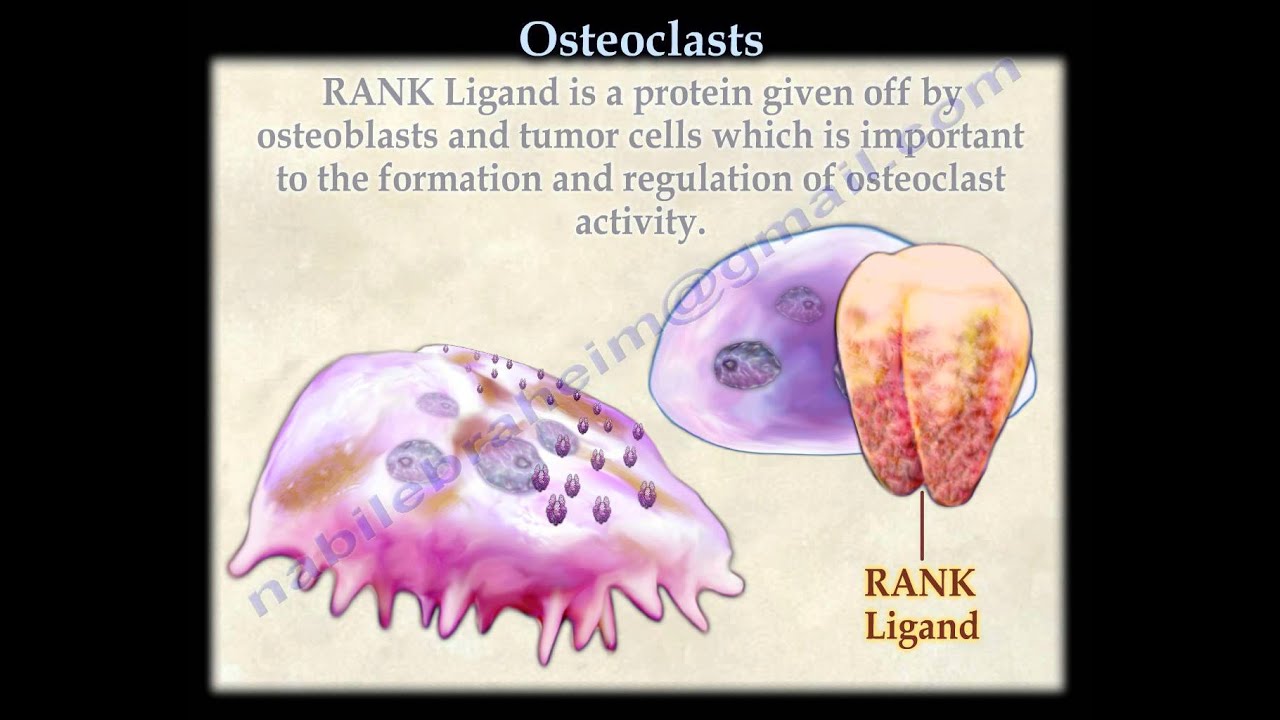
- Osteoblasts secrete important factors that activate osteoclasts, i.e., RANK ligand and other associated factors which communicate with other cells.
- Osteoblasts also secrete a regulatory protein involved in regulating phosphate excretion from the kidneys.
- After osteoblasts have finished making up new bone, some will differentiate into osteocytes, and the rest will surround the bone matrix. Some of them, if they remained, would either stay on the surface of the new bone or mature into lining cells.
- Lining cells are involved in the transport of calcium from inside and outside of the bone.
- Non-useful osteoblasts undergo the programmed cell suicide process (apoptosis) and will get disintegrated.
![]()
Osteocytes
History of Osteocytes
Osteocytes are the most abundant and long-lived bone cells, with speculation of living for about 25 years.
- During the 1950s, Gordan and Ham extensively studied osteocytes. In previous days of osteocyte discovery, it was thought that osteocytes were dormant cells and did not perform any function.
- With the passage of time and technological advancement, methods such as the isolation culturing of bone cells, the formation of phenotypically stable cell lines, and new animal models led to a better understanding of osteocytes.
![]()
Structure of Osteocytes
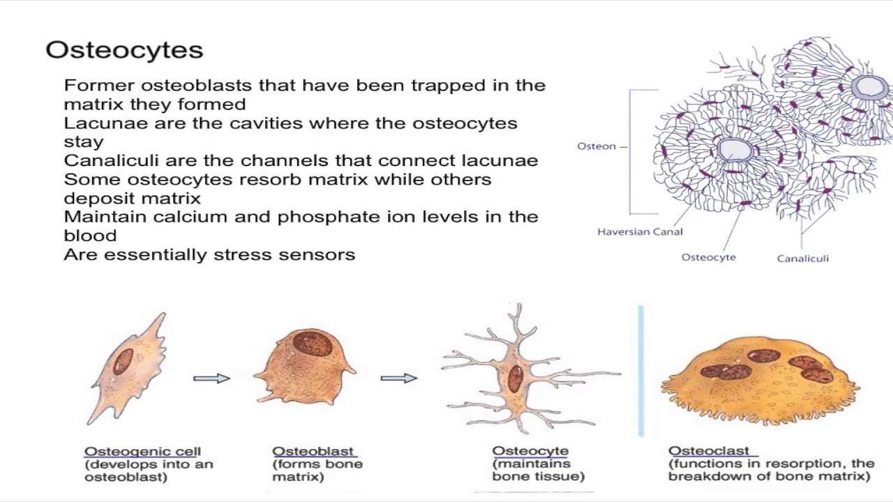
Osteocytes are located inside the bone and have a connection with each other and with other cells with the help of their long branches.
- Osteocytes are in the perfect position to sense any pressure or mechanical strain in the bone. It is estimated that osteocytes comprise about 95% of the total cells of the bone.
- Osteocytes are located in lacunae within the mineralized bone matrix. They possess a dendritic morphology.
- It was also observed that osteocytes might differ in morphology by the bone type in which they are present.
- Osteocytes with round shapes are present in the trabecular bones, and elongated osteocytes are in the cortical bone.
Osteocytes are derived from mesenchymal stem cells (MSCs) lineage through the Differentiation of osteoblasts. In this process, four stages have been proposed or recognized:
- Osteoid-osteocyte
- Pre-osteocyte
- Young osteocyte
- Mature osteocyte
The process of osteoblast differentiation is a tedious process involving many morphological and ultra-structural changes.
The most significant change is the reduction in the size of osteoblasts. Many major organelles decrease, but the size of the nucleus increases, corresponding to a decrease in the secretion and synthesis of proteins.
![]()
Function of Osteocytes
Initially, osteocytes were defined according to their morphology rather than their function. This was because their function remained unknown for decades. Later, it was recognized that they play many different yet important roles in bone development and maintenance.
Osteocytes perform different functions such as
- Osteocytes feature secreted growth factors that activate cells lining them or stimulate osteoblasts.
- The shape and arrangement of osteocytes help in the mechanical functioning of bones. The sensing ability and signal transportation characteristics also play a crucial role in this particular function. This phenomenon of osteocyte mechanical endurance occurs through piezoelectric effect.
- The functional bone adaptation is regulated by the osteocytes.
- The exchange of ions across the bone occurs with the help of osteocytes.
![]()
General Functions of Bone Cells
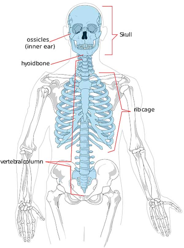
The bone cells perform diverse functions inside the human body. Some of the main functions of the bone cells are listed below:
- Osteoclasts maintain the ruffled borders in the bones.
- Formation of bone marrow occurs with the help of osteoclasts.
- Osteoclasts work under parathyroid hormone (PTH) to dissolve the bone.
- Osteoclasts produce factors known as “clastokines“, which influence the working of osteoblasts.
- Osteoclasts are also involved in regulating the hematopoietic stem cell niche.
- Osteoblasts are involved in the formation of the unmineralized osteoid part of the bone.
- Osteoblasts secrete enzymes known as alkaline phosphatase that create an acidic environment within their vicinity.
- Osteoblasts also produce hormones like prostaglandins that are fatty in nature.
- Osteoblasts have a role in the production of collagenous and non-collagenous proteins of the bone.
- Lining cells of bones help regulate the influx and the efflux of calcium ions from the bone.
- Lining cells also regulate the bone fluid via the regulation of mineral homeostasis.
- Lining cells help in the activation of osteoclasts indirectly.
- Osteocytes help in the bone turnover process and limit the dissolution of the bone.
- The bone adaptation function is performed by osteocytes coordinating the bone mechanical strength with the surrounding environment.
- Osteocytes play a crucial role in calcium homeostasis.
- Osteocytes help in the maintenance of bone matrix.
- Osteocytes have a role in sensing pressure or crack of the bone and signaling other parts of the bone.
- Upon mechanical stimulations, osteocytes produce secondary messengers such as adenosine triphosphate (ATP).
Despite the medical and technological advancements, the full functioning of the bone cells is yet to be elucidated.
![]()
The crucial aspect of the bone cells functioning would be to understand how the vast array of functions performed by bone cells can be used in the management of advanced bone dysfunctioning?
Recent scientific advancements and stem cell culturing techniques provide a beacon of light to unveil the hidden secrets of miraculous bone cells.
![]()
Cite this page
Bio Explorer. (2026, January 28). Types of Bone Cells. https://www.bioexplorer.net/types-of-bone-cells.html/

