
Immunology Methods and Protocols

Immunology protocols are practical laboratory methods used to study immune cells, antibodies, antigens, cytokines, inflammation, immune memory, immune signaling, vaccine responses, autoimmunity, allergy, infection, cancer immunity, and immune-related disease. These methods help researchers detect immune molecules, identify immune cell populations, measure cytokine release, test antibody binding, stain tissues or cells, analyze T cell responses, and connect immune data back to a biological question.
This page is a guide to major immunology methods and protocols used in research labs, teaching labs, clinical immunology, vaccine research, cancer immunology, infection biology, molecular biology, cell biology, microbiology, and biotechnology. It explains what each method is for, what a good protocol should include, where mistakes usually happen, and which trusted resources can help you go deeper.
A useful immunology protocol is more than a list of steps. It should explain the sample type, immune target, antigen or antibody, controls, reagents, instrument settings, biosafety requirements, data readout, gating or analysis plan, troubleshooting, and limitations. Nature Protocols describes strong protocols as including reagents, equipment, timing, procedures, design advice, limitations, troubleshooting, data analysis, and result interpretation.
The Adjuvant Mixing Calculator flags unsafe species-route-adjuvant combinations against NIH OACU per-site volume limits before you mix a batch.
What Are Immunology Protocols?
Immunology protocols are written workflows for measuring immune cells, immune proteins, immune reactions, and immune function under defined conditions. They may be used to detect an antibody, quantify a cytokine, identify a T cell subset, stain an immune marker, measure antigen-specific responses, test complement activity, analyze inflammation, or compare immune responses before and after treatment.
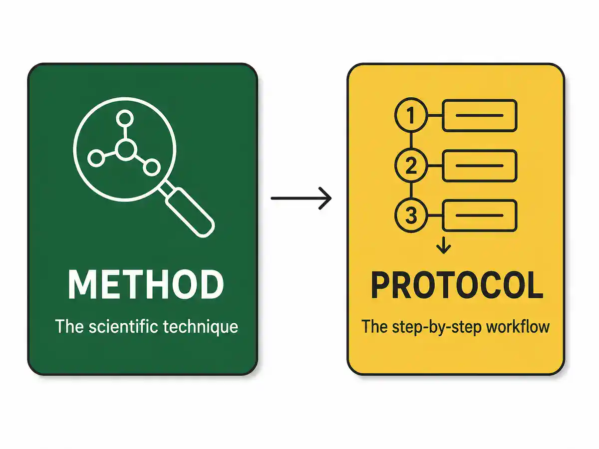
The method is the scientific technique. The protocol is the practical workflow for using that technique. For example, ELISA is a method for detecting or quantifying an antigen or antibody. An ELISA protocol describes the sample, plate format, coating or capture strategy, blocking step, standards, detection antibody, enzyme substrate, controls, reading wavelength, standard curve, and interpretation rules.
Immunology overlaps closely with immunology, cell biology, molecular biology, microbiology, biochemistry, virology, biotechnology, pharmacology, pathology, oncology, vaccine science, and clinical medicine.
Immunology Protocols Guide
Use this page as a map for common immunology lab methods and immune assay workflows.
- biosafety and immune sample handling
- antibody selection and antibody validation
- ELISA and immunoassays
- flow cytometry and immunophenotyping
- intracellular cytokine staining
- ELISpot and FluoroSpot assays
- immunofluorescence and immunocytochemistry
- immunohistochemistry
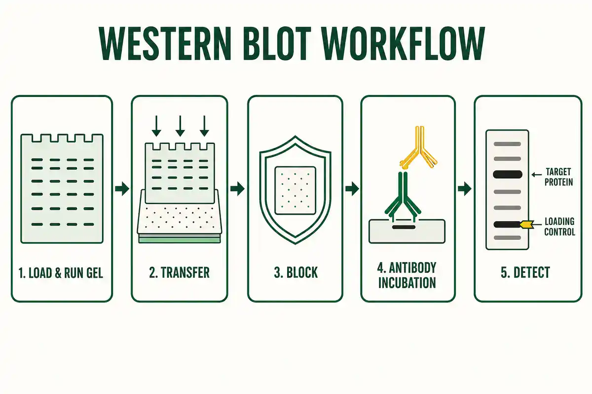
- western blotting for immune proteins
- cytokine and chemokine assays
- immune cell isolation and PBMC workflows
- complement assays and antibody function assays
- vaccine immunogenicity and immune monitoring
- controls, troubleshooting, reporting, and quality checks
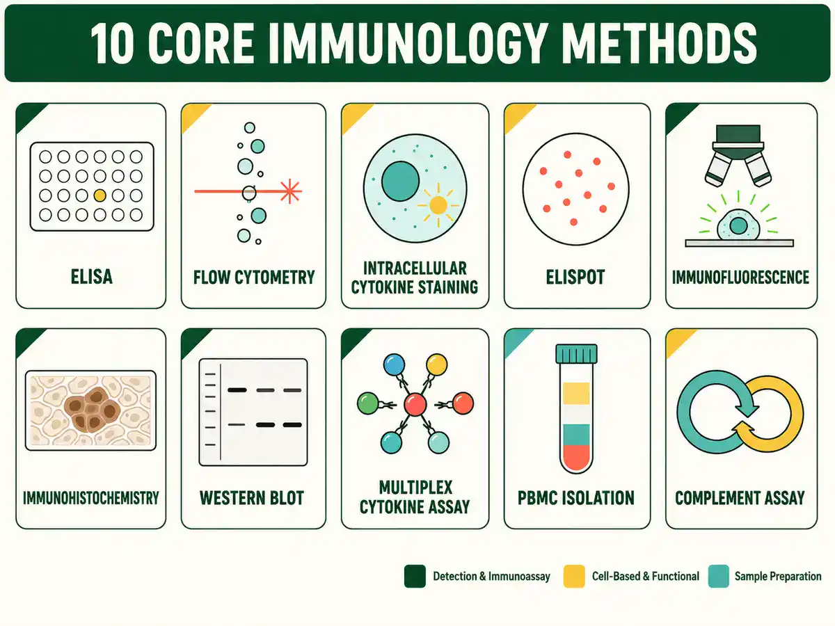
Core Immunology Methods at a Glance
The table below gives a quick map of important immunology methods and what each one helps measure or interpret.
| Method | Main Purpose | Common Readout |
|---|---|---|
| ELISA | Detect or quantify antibodies, antigens, cytokines, hormones, or immune proteins | Absorbance, standard curve, concentration, positive or negative result |
| Flow cytometry | Measure markers on individual cells in suspension | Cell populations, fluorescence intensity, gating plots, percent positive cells |
| Intracellular cytokine staining | Detect cytokines inside stimulated immune cells | Cytokine-positive T cells, marker combinations, functional cell subsets |
| ELISpot | Detect individual cells secreting a cytokine or antibody | Spot count, spot-forming cells, antigen-specific response |
| Immunofluorescence | Visualize immune markers in cells or tissues using fluorescent antibodies | Fluorescent staining pattern, localization, intensity, colocalization |
| Immunohistochemistry | Detect antigens in tissue sections using antibody-based staining | Positive cells, tissue localization, staining intensity, pathology context |
| Western blot | Detect a specific immune-related protein after gel separation | Band size, band intensity, loading control, specificity controls |
| Multiplex cytokine assay | Measure multiple cytokines or chemokines in one sample | Concentration panel, cytokine pattern, inflammatory profile |
| PBMC isolation | Separate peripheral blood mononuclear cells for immune assays | Cell yield, viability, lymphocyte and monocyte fraction, downstream assay quality |
| Complement assay | Measure complement activity or complement pathway function | Hemolysis, deposition signal, pathway activity, inhibition response |
Biosafety and Immune Sample Handling
Immunology protocols often begin with human blood, serum, plasma, tissue, primary immune cells, animal samples, infectious material, recombinant proteins, viral vectors, or genetically modified cells. These materials can carry biological, chemical, physical, and regulatory risks.
The CDC describes biological risk assessment as a continuous risk management process that identifies hazards, evaluates risks, applies controls, and monitors whether the controls remain effective. The assessment should consider the biological agent, procedure, facility, equipment, and worker competency. See CDC Biological Risk Assessment.
A strong immunology protocol should state the sample source, biosafety level, PPE, specimen handling conditions, decontamination method, sharps policy, waste disposal, centrifugation precautions, aerosol risks, transport conditions, and whether institutional approval or ethics approval is required. For human-derived material, local biosafety and privacy rules matter as much as the assay chemistry.
This page is educational. It does not replace institutional SOPs, biosafety committee guidance, human-subjects approval, animal-care approval, clinical laboratory rules, or supervisor training.
Antibody Selection and Antibody Validation
Many immunology methods depend on antibodies. Antibodies are powerful because they can bind specific antigens, but that specificity must be tested in the actual application. An antibody that works in western blotting may not work in flow cytometry. An antibody that works in fixed tissue may not work in live-cell staining. An antibody that gives a beautiful image may still be nonspecific without proper controls.
A Nature Methods proposal for antibody validation recommends using multiple independent strategies to test antibody specificity, including genetic, orthogonal, independent-antibody, tagged-protein, and immunocapture approaches. See A proposal for validation of antibodies.
A useful antibody-based protocol should document the target, clone, host species, isotype, conjugate, lot number, application, dilution or titration, sample type, fixation or permeabilization conditions, positive control, negative control, secondary antibody, blocking strategy, and expected staining pattern. If the antibody is used for flow cytometry, the protocol should also explain compensation, fluorescence-minus-one controls, and gating logic.
The simplest rule is: do not trust an antibody because the label says it is specific. Trust it when the evidence matches the sample, application, controls, and biological question.
ELISA and Immunoassay Protocols
ELISA protocols, short for enzyme-linked immunosorbent assay protocols, are among the most common immunology lab methods. ELISA can detect or quantify antibodies, antigens, cytokines, hormones, peptides, proteins, immune complexes, and other analytes using antigen-antibody recognition and enzyme-based signal detection.
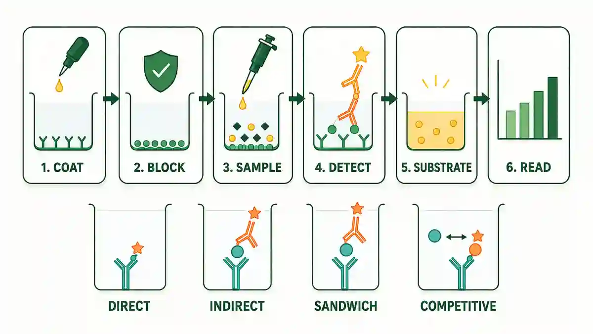
NCBI Bookshelf describes ELISA as a heterogeneous enzyme immunoassay in which a reaction component is immobilized on a solid phase such as a microtiter well, magnetic particle, or plastic bead. See Enzyme-Linked Immunosorbent Assay. Thermo Fisher's ELISA guide explains that ELISA formats depend on common elements such as coating or capture, blocking, detection, washing, and signal development. See ELISA Technical Guide and Protocols.
Common ELISA formats include direct ELISA, indirect ELISA, sandwich ELISA, competitive ELISA, and multiplex immunoassays. The best format depends on the target, sample complexity, antibody pair, expected concentration range, sensitivity requirement, available standards, and whether the goal is detection or quantification.
A useful ELISA protocol should state the plate type, capture antigen or antibody, blocking reagent, sample dilution, standards, controls, incubation conditions, wash buffer, detection antibody, enzyme conjugate, substrate, stop solution, plate reader wavelength, standard curve model, replicate strategy, and acceptance rules.
Common ELISA mistakes include weak standard curves, plate edge effects, poor washing, wrong sample dilution, high background, expired substrate, cross-reactive antibodies, inconsistent incubation time, and treating values outside the standard curve as reliable. A good ELISA page should teach the reader how to judge the result, not only how to produce color in a well.
Flow Cytometry and Immunophenotyping Protocols
Flow cytometry protocols measure physical and fluorescent properties of individual cells in a flowing sample. In immunology, flow cytometry is widely used for immunophenotyping, lymphocyte subset analysis, cytokine detection, cell activation, apoptosis, cell cycle, proliferation, viability, immune monitoring, and cell sorting.
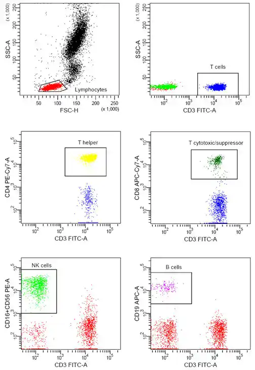
NCBI Bookshelf describes flow cytometry as a technique that analyzes light scattering and fluorescence properties of cells. A review of flow cytometry explains that lasers generate scattered and fluorescent signals that are read by detectors. See Flow Cytometry Blood Cell Identification and Flow Cytometry: An Overview.
A useful flow cytometry protocol should state the sample type, cell preparation method, viability dye, antibody panel, fluorophores, staining buffer, fixation conditions if used, compensation controls, fluorescence-minus-one controls, isotype controls when appropriate, gating strategy, instrument configuration, acquisition threshold, event count, data transformation, and analysis software.
The MIFlowCyt standard was created to define the minimum information needed to interpret flow cytometry experiments, including specimen details, reagents, instrument configuration, and data processing. See MIFlowCyt: The Minimum Information about a Flow Cytometry Experiment.
Flow cytometry is not just "stain cells and read the machine." The quality of the result depends on sample health, cell clumping, antibody titration, marker choice, spillover, compensation, gating, autofluorescence, dead-cell exclusion, and whether the cell population being analyzed makes biological sense.
Intracellular Cytokine Staining Protocols
Intracellular cytokine staining, often shortened to ICS, is used to detect cytokines inside immune cells after stimulation. It is especially useful for studying T cell responses, antigen-specific immunity, vaccine responses, infection, inflammation, and immune monitoring.
An ICS protocol usually needs more planning than surface-marker staining because it combines stimulation, secretion blocking, surface staining, fixation, permeabilization, intracellular staining, flow cytometry acquisition, and gating. The protocol should clearly separate surface markers from intracellular cytokine markers and should preserve both cell identity and cytokine signal.
MIATA, or Minimal Information About T Cell Assays, was developed to improve reporting of T cell assay results and includes immune monitoring methods such as intracellular cytokine staining, ELISpot, and HLA-peptide multimer staining. See MIATA: Minimal Information About T Cell Assays.
A useful ICS protocol should state the cell source, antigen or stimulant, stimulation time, secretion inhibitor, surface markers, viability dye, fixation and permeabilization reagents, cytokine antibodies, positive control, negative control, compensation controls, fluorescence-minus-one controls, gating plan, and reporting format.
Common ICS mistakes include overstimulation, poor cell viability, weak positive controls, missing unstimulated controls, poor fixation-permeabilization compatibility, marker loss after fixation, and gating cytokine-positive cells without checking background signal.
Cell Viability Dose-Response Calculator
ELISpot and FluoroSpot Assay Protocols
ELISpot protocols detect individual cells that secrete a target molecule, commonly a cytokine such as interferon-gamma. FluoroSpot extends this idea by using fluorescent detection to measure more than one secreted molecule in the same well.
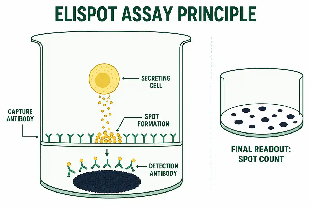
ELISpot is often used in vaccine research, T cell immune monitoring, infection studies, cancer immunology, transplantation research, and gene therapy immune-response monitoring. The readout is usually a spot count, often reported as spot-forming cells after background subtraction and normalization.
A useful ELISpot protocol should state the cell type, cell number per well, antigen or stimulant, positive control, negative control, capture antibody, detection antibody, incubation time, plate type, spot development method, counting method, background rule, replicate strategy, and acceptance criteria.
ELISpot is sensitive, but the result can be affected by cell viability, peptide quality, stimulation time, high background, plate handling, spot saturation, inconsistent washing, and subjective spot counting. Good protocols define how borderline wells, merged spots, high background, and outlier replicates will be handled before the experiment is interpreted.
Immunofluorescence and Immunocytochemistry Protocols
Immunofluorescence protocols use antibodies linked directly or indirectly to fluorophores to visualize proteins, immune markers, organelles, or cellular structures. In immunology, immunofluorescence can show where immune markers are located in cells or tissues and how localization changes after stimulation, infection, treatment, or differentiation.
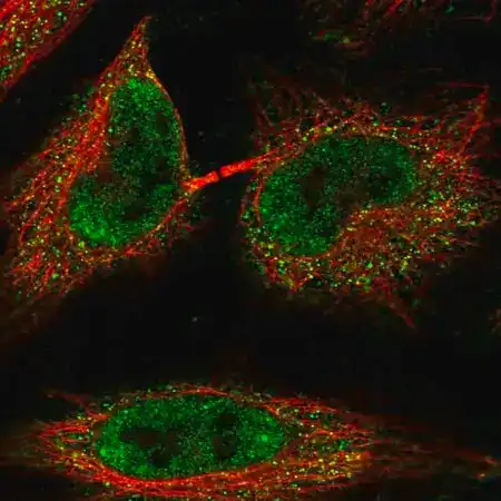
(Huaijin2002, CC BY-SA 4.0
Abcam describes immunocytochemistry, also known as immunofluorescence, as a method for visualizing the localization and distribution of proteins of interest within cultured cells, with steps such as fixation, permeabilization, blocking, primary antibody staining, secondary antibody staining, and counterstaining. See Immunocytochemistry Protocol.
A useful immunofluorescence protocol should state the cell or tissue type, fixation method, permeabilization method, blocking reagent, primary antibody, secondary antibody, counterstain, mounting medium, microscope type, exposure settings, positive control, negative control, secondary-only control, and image-analysis method.
Common immunofluorescence mistakes include over-fixation, weak permeabilization, high background, photobleaching, cross-reactive secondary antibodies, poor antibody validation, inconsistent imaging settings, and overprocessing images after acquisition.
Immunohistochemistry Protocols
Immunohistochemistry protocols, often shortened to IHC, detect antigens in tissue sections using antibody-based staining. IHC is widely used in pathology, cancer biology, infection biology, neuroscience, developmental biology, and tissue immunology.
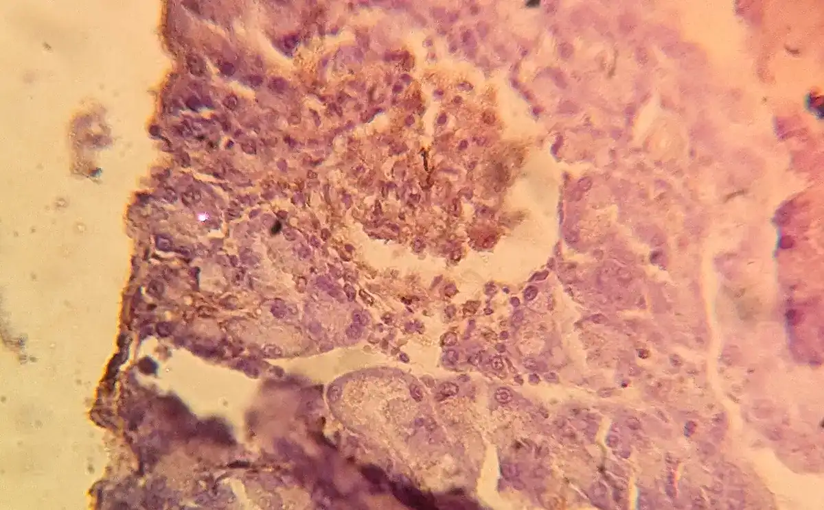
IHC protocols must preserve tissue architecture while exposing the antigen to antibody detection. This balance can be difficult because fixation, embedding, sectioning, antigen retrieval, blocking, antibody choice, detection chemistry, and counterstaining all affect the final signal.
A useful IHC protocol should state the tissue type, fixation method, section thickness, antigen retrieval method, blocking step, primary antibody, detection system, chromogen or fluorophore, counterstain, control tissue, negative control, scoring method, and whether interpretation is qualitative, semi-quantitative, or image-analysis based.
A brown stain or fluorescent signal is not automatically a correct result. Tissue autofluorescence, endogenous enzyme activity, nonspecific binding, necrotic tissue, edge artifacts, and poor antibody specificity can all mislead interpretation.
Cytokine and Chemokine Assay Protocols
Cytokine assay protocols measure immune signaling molecules such as interleukins, interferons, tumor necrosis factor, chemokines, colony-stimulating factors, and inflammatory mediators. These assays are common in infection, vaccine, allergy, autoimmune, cancer, sepsis, transplant, and inflammation research.
Cytokines can be measured by ELISA, multiplex bead assays, intracellular cytokine staining, ELISpot, qPCR, protein arrays, or functional bioassays. These methods do not answer the same question. ELISA measures soluble protein in a sample. ICS asks which cells contain a cytokine after stimulation. ELISpot estimates secreting cells. qPCR measures transcript abundance, not secreted protein.
A useful cytokine assay protocol should state sample type, collection time, storage conditions, freeze-thaw history, anticoagulant if relevant, stimulation conditions, standard curve, assay range, dilution plan, controls, replicate strategy, normalization method, and whether results are reported as concentration, fold change, percent positive cells, or spot-forming cells.
Cytokine data are easy to overinterpret. A single cytokine rarely explains an immune response by itself. Timing, sample type, cell source, stimulation method, disease context, and assay platform all shape the result.
PBMC Isolation and Immune Cell Preparation
PBMC isolation protocols separate peripheral blood mononuclear cells from blood. PBMCs include lymphocytes and monocytes and are widely used for flow cytometry, ELISpot, T cell assays, cytokine studies, vaccine immunogenicity testing, transcriptomics, and immune monitoring.
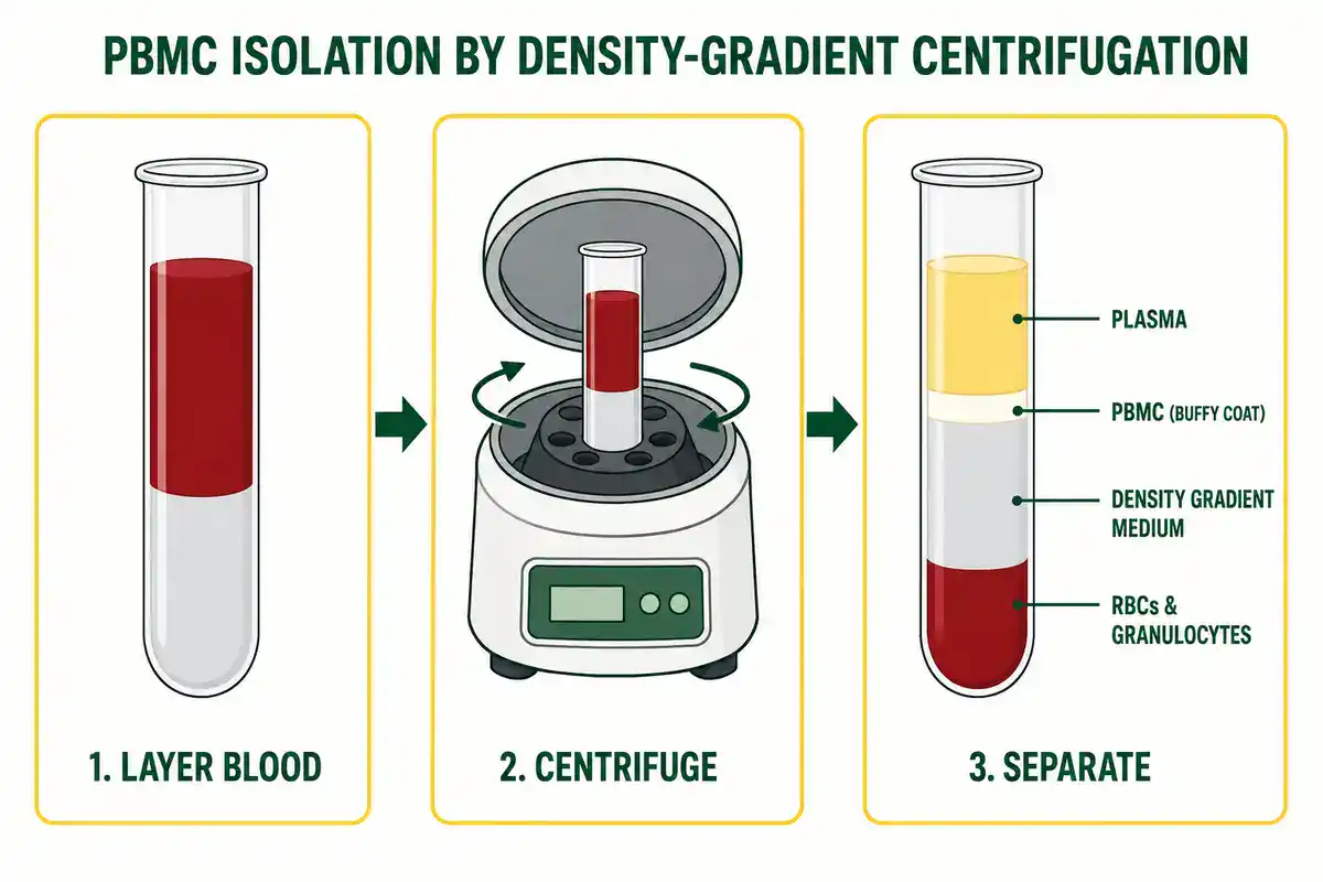
A useful immune cell preparation protocol should state blood source, anticoagulant, processing time, separation method, washing conditions, cell count, viability, platelet removal if relevant, red-cell contamination rule, cryopreservation method, thawing method, resting time, and downstream assay requirements.
PBMC quality can change quickly. Delayed processing, rough handling, temperature changes, red-cell contamination, platelet carryover, low viability, and freeze-thaw stress can all affect immune assays. For longitudinal studies, the protocol should keep sample handling as consistent as possible across time points.
Antibody Function and Complement Assays
Some immunology protocols ask not only whether an antibody binds, but what the antibody does. Functional assays may measure neutralization, opsonophagocytosis, antibody-dependent cellular cytotoxicity, complement activation, receptor blocking, or immune-complex formation.
Complement assay protocols measure activity in complement pathways or detect complement proteins and activation products. These assays may be used in immunology research, autoimmune disease studies, infection biology, vaccine evaluation, and clinical immunology.
A useful antibody function or complement protocol should state the biological target, sample source, heat-inactivation status if relevant, cell type or target system, serum or plasma handling, controls, reference standard, endpoint, pathway being tested, and whether the assay measures binding, activity, inhibition response, deposition, or cell killing.
Binding and function are not the same. An antibody may bind strongly but fail to neutralize. Another antibody may bind weakly but still trigger a functional pathway under the right conditions. Good protocols make the measured function explicit.
Immune Monitoring and Vaccine Response Protocols
Immune monitoring protocols measure how an immune system responds across time, conditions, treatments, vaccines, infections, tumors, transplants, or therapies. These workflows may combine flow cytometry, ELISA, ELISpot, cytokine panels, neutralization assays, sequencing, transcriptomics, and clinical metadata.
A useful immune monitoring protocol should state the study time points, sample type, collection method, processing time, storage conditions, assay platform, antigen or stimulation condition, positive and negative controls, reference materials, batch strategy, data normalization, and reporting standard.
MIATA was created because T cell assay data are hard to compare when important details are missing. The MIATA framework aims to provide the minimum information needed to understand how T cell assay data were generated and interpreted. See Minimal information about T cell assays: the process of reaching the community of T cell immunologists in cancer and beyond.
For BioExplorer, this section is a natural place for future sub-pages on vaccine immune-response assays, T cell assays, antibody titer interpretation, neutralization assay principles, and immune monitoring workflows.
How to Choose the Right Immunology Protocol
Do not choose an immunology protocol only because it appears first in search results. Choose it because it matches your immune target, sample type, assay goal, safety level, instrument, and analysis plan.
Before using any protocol, check these points:
- Sample type: serum, plasma, whole blood, PBMCs, tissue, cultured immune cells, supernatant, bronchoalveolar lavage, lymphoid organ, tumor sample, or animal sample.
- Immune target: antibody, antigen, cytokine, chemokine, cell-surface marker, transcription factor, intracellular protein, complement protein, or immune cell subset.
- Goal: detection, quantification, localization, immunophenotyping, functional response, immune monitoring, vaccine response, or assay validation.
- Assay format: ELISA, flow cytometry, ELISpot, immunofluorescence, immunohistochemistry, western blot, multiplex bead assay, neutralization assay, or complement assay.
- Controls: positive control, negative control, unstimulated control, vehicle control, isotype control, secondary-only control, fluorescence-minus-one control, compensation control, standard curve, or reference sample.
- Antibody quality: clone, lot, host species, isotype, conjugate, application validation, titration, specificity evidence, and storage history.
- Instrument settings: plate reader wavelength, microscope exposure, flow cytometer lasers and detectors, compensation, threshold, event count, and acquisition template.
- Safety: human material, animal material, infectious risk, recombinant material, chemical hazards, sharps, aerosols, and waste disposal.
- Verification: know how success will be checked before beginning the protocol.
Common Mistakes in Immunology Protocols
Immunology methods often fail for ordinary reasons. The assay may be valid, but the sample, antibody, control design, or analysis plan may not fit the question.
- Using unvalidated antibodies: antibody specificity must be tested for the actual application, sample, and species.
- Skipping antibody titration: too much antibody can raise background, while too little can hide real signal.
- Ignoring sample handling: delayed processing, freeze-thaw cycles, poor viability, or hemolysis can distort immune readouts.
- Weak controls: missing positive, negative, unstimulated, compensation, or fluorescence-minus-one controls makes interpretation fragile.
- Overinterpreting ELISA values: readings outside the standard curve range should not be treated as reliable concentrations.
- Bad flow cytometry gates: poor gating can create or erase cell populations.
- Confusing mRNA and protein: qPCR cytokine transcripts do not always match secreted cytokine protein levels.
- Comparing different assay platforms as if they are identical: ELISA, ELISpot, ICS, and multiplex assays answer related but different questions.
- Batch effects: plate, reagent lot, operator, instrument, and day-to-day differences can affect immune assay results.
- Publishing incomplete methods: missing sample, antibody, instrument, gating, or analysis details can make immune data hard to interpret or reproduce.
Immunology Calculators and Lab Tools
Immunology protocols often depend on calculations. A wrong sample dilution, antibody dilution, standard curve, cytokine concentration, cell recovery estimate, gating percentage, or spot-forming-cell calculation can change the result before interpretation begins.
Useful immunology calculators include:
- ELISA standard curve calculator
- antibody dilution calculator
- serial dilution calculator
- cytokine concentration calculator
- ELISpot spot-forming-cell calculator
- flow cytometry percentage calculator
- gating population frequency calculator
- stain index calculator
- compensation spillover helper
- PBMC recovery calculator
- cell viability calculator
- antibody titer calculator
- neutralization IC50 calculator
- multiplex cytokine normalization calculator
- plate layout calculator
BioExplorer's Biology Tools and Calculators hub is building free browser-based tools by branch of biology. The existing Genetics and Inheritance Tools section includes inheritance and population genetics tools, while the Botany Tools section supports plant science workflows.
As BioExplorer expands its immunology tools, this page can link directly to ELISA calculators, antibody dilution tools, immune-cell analysis helpers, and cytokine assay calculators.
Trusted Immunology Protocol Resources
Use BioExplorer as a guide, but always check your institution's approved SOPs, biosafety rules, manufacturer instructions, ethics approvals, and supervisor guidance before performing real laboratory work. These external resources are useful starting points for immunology protocols and method background:
- Nature Protocols: peer-reviewed protocol articles with design advice, troubleshooting, data analysis, limitations, and result interpretation.
- CDC Biological Risk Assessment: guidance on identifying hazards, assessing risk, applying controls, and monitoring effectiveness.
- NCBI Bookshelf: Enzyme-Linked Immunosorbent Assay: background on ELISA and enzyme immunoassay principles.
- Thermo Fisher ELISA Technical Guide: ELISA formats, coating, blocking, washing, detection, and signal development concepts.
- Flow Cytometry: An Overview: review of flow cytometry principles, lasers, scattered light, fluorescence, and detectors.
- MIFlowCyt Reporting Standard: minimum information needed to interpret flow cytometry experiments.
- MIATA: Minimal Information About T Cell Assays: reporting framework for T cell immune monitoring assays, including ICS, ELISpot, and HLA-peptide multimer staining.
- Abcam Immunocytochemistry Protocol: immunofluorescence workflow covering fixation, permeabilization, blocking, antibody staining, and counterstaining.
- A proposal for validation of antibodies: Nature Methods antibody validation framework using multiple independent strategies.
- Bio-Rad FMO Controls for Flow Cytometry: explanation of fluorescence-minus-one controls for setting gates in multicolor flow cytometry panels.
Safety and Responsibility
Immunology protocols can involve human blood, primary cells, animal tissue, infectious agents, recombinant proteins, viral vectors, cytokines, adjuvants, antibodies, chemical fixatives, fluorescent dyes, sharps, centrifuges, biosafety cabinets, and regulated biological waste. This page is educational. It does not replace formal training, institutional SOPs, biosafety approval, chemical safety guidance, ethics approval, or supervision by qualified personnel.
Do not handle human-derived samples, infectious material, recombinant organisms, viral vectors, animal tissues, or regulated biological materials without the proper facility, training, approvals, containment, and disposal procedures. When in doubt, stop and follow institutional biosafety and ethics guidance.
Related BioExplorer Resources
- Biology Methods and Protocols: main hub for biology lab methods, protocol categories, safety notes, and trusted resources.
- Immunology: guide to immune cells, antibodies, antigens, inflammation, immunity, and immune-system function.
- Immunology Glossary: clear definitions of antibodies, antigens, cytokines, lymphocytes, inflammation, hypersensitivity, immune memory, and related terms.
- Cell Biology Methods and Protocols: methods for cell culture, cell counting, viability, staining, microscopy, flow cytometry, and cell-based assays.
- Molecular Biology Methods and Protocols: protocols for DNA, RNA, PCR, cloning, gel electrophoresis, sequencing prep, and molecular analysis.
- Microbiology Methods and Protocols: methods for aseptic technique, microbial culture, Gram staining, serial dilution, CFU counting, antimicrobial testing, and biofilms.
- Biochemistry Methods and Protocols: methods for protein assays, SDS-PAGE, western blotting, enzyme kinetics, chromatography, and protein purification.
- Biology Tools and Calculators: free browser-based biology calculators and educational tools.
Frequently Asked Questions
Immunology protocols are written laboratory workflows for studying immune cells, antibodies, antigens, cytokines, inflammation, immune responses, and immune-related disease under defined conditions.
Common immunology protocols include ELISA, flow cytometry, immunophenotyping, intracellular cytokine staining, ELISpot, immunofluorescence, immunohistochemistry, western blotting, cytokine assays, PBMC isolation, and antibody validation workflows.
ELISA is used to detect or quantify antibodies, antigens, cytokines, hormones, immune proteins, and other analytes using antigen-antibody binding and enzyme-based signal detection.
Controls help separate true biological signal from background, autofluorescence, spectral spillover, dead cells, nonspecific binding, and gating errors. Compensation controls and fluorescence-minus-one controls are especially important in multicolor panels.
Intracellular cytokine staining is a flow cytometry method used to detect cytokines inside immune cells after stimulation. It helps identify which cell types are producing cytokines under defined conditions.
ELISA usually measures the amount of a soluble target in a sample. ELISpot detects individual cells that secrete a target molecule, often reported as spot-forming cells.
Antibody validation matters because nonspecific or poorly matched antibodies can produce misleading signals. Validation should match the sample, species, application, controls, and expected biological pattern.
Not always. Online protocols vary in quality and may not match your sample, biosafety level, instrument, antibody, cell type, or institution's rules. For real lab work, follow approved SOPs, safety guidance, manufacturer instructions, ethics approval, and supervisor training.
Cite this page
BioExplorer. (2026, August 26). Immunology Methods and Protocols. https://www.bioexplorer.net/methods_and_protocols/immunology/