
Biophysics Methods and Protocols

Biophysics protocols are practical laboratory and computational methods used to study biological systems with the tools of physics, chemistry, mathematics, engineering, and quantitative biology.
These methods help researchers measure molecular structure, binding affinity, reaction kinetics, thermodynamics, membrane behavior, protein folding, molecular motion, cell mechanics, ion flow, fluorescence signals, and force at biological scales.
Biophysics Methods and Protocols Guide:
- What Are Biophysics Protocols?
- Biophysics Protocols Guide
- Core Biophysics Methods at a Glance
- Sample Preparation for Biophysical Methods
- Structural Biophysics: X-ray, NMR and Cryo-EM
- Small-Angle Scattering and Integrative Structural Methods
- Circular Dichroism and Protein Folding Protocols
- Fluorescence Spectroscopy and FRET Protocols
- Surface Plasmon Resonance and Binding Kinetics
- Isothermal Titration Calorimetry and Binding Thermodynamics
- Differential Scanning Calorimetry and Thermal Stability
- Dynamic Light Scattering and SEC-MALS
- Atomic Force Microscopy and Force Spectroscopy
- Optical Tweezers and Single-Molecule Force Methods
- Electrophysiology and Membrane Biophysics
- Molecular Dynamics and Computational Biophysics
- How to Choose the Right Biophysics Protocol
- Common Mistakes in Biophysics Protocols
- Biophysics Calculators and Lab Tools
- Trusted Biophysics Protocol Resources
- Safety and Responsibility
- Related BioExplorer Resources
- Frequently Asked Questions
This page is a guide to major biophysics methods and protocols used in molecular biophysics, structural biology, membrane biophysics, protein science, cell biophysics, electrophysiology, drug discovery, biochemistry, molecular biology, computational biology, and biomedical research.
It explains what each method is for, what a good protocol should include, where mistakes usually happen, and which trusted resources can help you go deeper.
A useful biophysics protocol is more than a list of instrument steps. It should explain the biological question, sample state, buffer, concentration range, controls, calibration, physical model, instrument settings, data quality checks, fitting assumptions, error sources, limitations, and interpretation. Nature Protocols emphasizes that strong protocols should add practical detail on experimental design, troubleshooting, data analysis, limitations, and result interpretation.
What Are Biophysics Protocols?
Biophysics protocols are written workflows for measuring how biological molecules, cells, membranes, and systems behave under physical laws. They may be used to determine a protein structure, quantify protein-ligand binding, measure membrane potential, track molecular motion, estimate diffusion, analyze protein stability, record ion-channel currents, or simulate molecular dynamics.
The method is the scientific technique. The protocol is the practical workflow for using that technique. For example, surface plasmon resonance is a method for measuring molecular interactions in real time. An SPR protocol describes ligand immobilization, analyte concentration series, flow rate, reference surface, regeneration condition, sensorgram processing, kinetic model, and binding interpretation.
Biophysics overlaps closely with biochemistry methods, molecular biology methods, cell biology methods, genetics methods, structural biology, pharmacology, neuroscience, computational biology, nanobiology, and bioengineering.
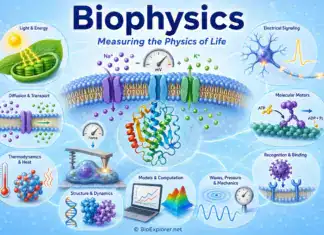
What Is Biophysics?
Biophysics Protocols Guide
Use this page as a map for common biophysical methods and quantitative biology workflows.
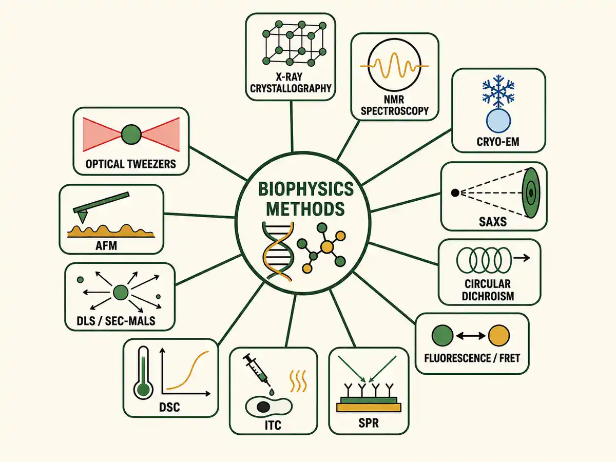
- protein and macromolecule sample preparation
- X-ray crystallography
- nuclear magnetic resonance spectroscopy
- cryo-electron microscopy
- small-angle X-ray scattering
- circular dichroism spectroscopy
- fluorescence spectroscopy and FRET
- surface plasmon resonance
- isothermal titration calorimetry
- differential scanning calorimetry
- dynamic light scattering
- SEC-MALS and molecular-weight analysis
- atomic force microscopy and force spectroscopy
- optical tweezers and single-molecule force methods
- patch clamp and electrophysiology
- membrane biophysics and transport measurements
- molecular dynamics simulations
- controls, fitting models, error analysis, and troubleshooting
Core Biophysics Methods at a Glance
The table below gives a practical overview of major biophysics methods and the kind of biological question each one helps answer.
| Biophysics Method | Main Purpose | Common Readout |
|---|---|---|
| X-ray crystallography | Determine atomic or near-atomic structures from ordered crystals | Electron density map, atomic model, resolution, R factor, ligand position |
| NMR spectroscopy | Study structure, dynamics, and interactions in solution | Chemical shifts, restraints, relaxation data, structural ensemble, binding perturbation |
| Cryo-EM | Determine structures of large complexes, particles, and conformational states | Electron density map, particle classes, 3D reconstruction, resolution estimate |
| SAXS | Measure low-resolution size and shape of macromolecules in solution | Radius of gyration, pair-distance distribution, envelope, oligomeric state clues |
| Circular dichroism | Estimate protein secondary structure and folding stability | CD spectrum, melting curve, thermal transition, folding change |
| Fluorescence and FRET | Measure molecular environment, distance changes, conformational shifts, or interactions | Emission spectra, FRET efficiency, anisotropy, lifetime, intensity change |
| SPR | Measure real-time molecular binding without a label | Sensorgram, association rate, dissociation rate, affinity, specificity |
| ITC | Measure binding thermodynamics directly in solution | KD, stoichiometry, enthalpy, entropy, binding curve |
| DLS | Estimate particle size and aggregation in solution | Hydrodynamic radius, polydispersity, size distribution, aggregation signal |
| AFM | Image surfaces and measure nanoscale mechanical properties | Topography, force curve, stiffness, adhesion, indentation, membrane mechanics |
| Patch clamp | Measure ion-channel and membrane electrical activity | Current, voltage, conductance, channel opening, action potential, membrane properties |
| Molecular dynamics | Simulate atomic motions and interactions over time | Trajectory, RMSD, RMSF, interaction energies, conformational changes |
Sample Preparation for Biophysical Methods
Biophysics often fails at the sample stage. Many methods require purified, stable, homogeneous, correctly folded, and properly buffered samples. A beautiful instrument cannot rescue a degraded protein, aggregated complex, mismatched buffer, unstable membrane preparation, or poorly characterized ligand.
A useful sample-preparation protocol should state the biological source, expression system if relevant, purification method, buffer composition, pH, salt concentration, reducing agent, detergent or lipid system, protein concentration, ligand concentration, storage condition, freeze-thaw history, and quality checks. For structural methods, purity and homogeneity often matter as much as concentration.
Common quality checks include SDS-PAGE, western blot, UV absorbance, fluorescence-based concentration measurement, mass spectrometry, size-exclusion chromatography, dynamic light scattering, activity assay, thermal shift assay, or analytical ultracentrifugation. The best check depends on the molecule and method.
See BioExplorer's Biochemistry Methods and Protocols page, which covers protein assays, SDS-PAGE, western blotting, chromatography, enzyme assays, and protein purification.
Structural Biophysics: X-ray, NMR and Cryo-EM
Structural biophysics protocols help reveal the shapes of proteins, nucleic acids, membranes, viruses, ribosomes, molecular machines, and biomolecular complexes. The major structure-determination methods include X-ray crystallography, nuclear magnetic resonance spectroscopy, and electron microscopy. PDB-101 explains that protein structures are commonly determined by X-ray crystallography, NMR spectroscopy, and electron microscopy, and that other biophysical techniques can also support integrative modeling. See PDB-101 Methods for Determining Structure.
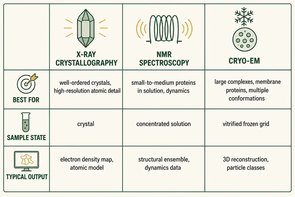
X-ray crystallography works best when the target can form well-ordered crystals. A crystallography protocol should describe protein purity, crystallization screen, crystal optimization, cryoprotection, diffraction collection, phasing strategy, model building, refinement, validation, and data-deposition details.
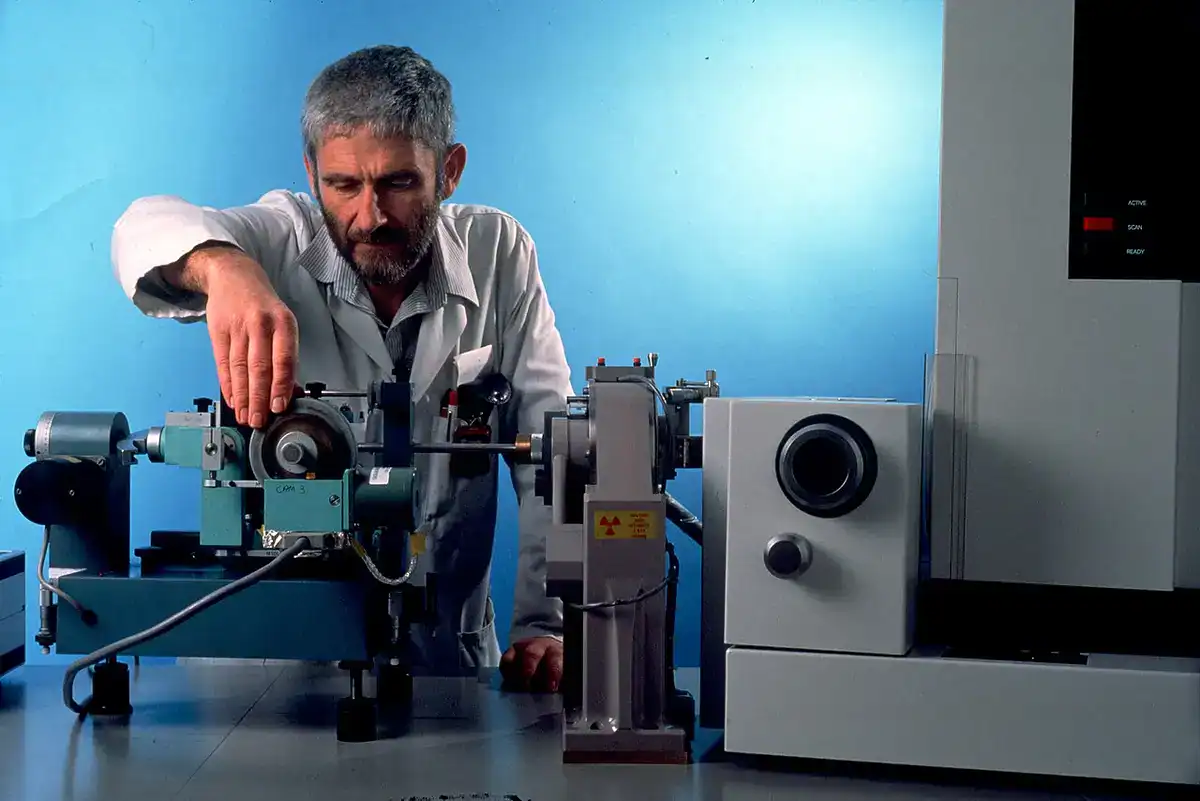
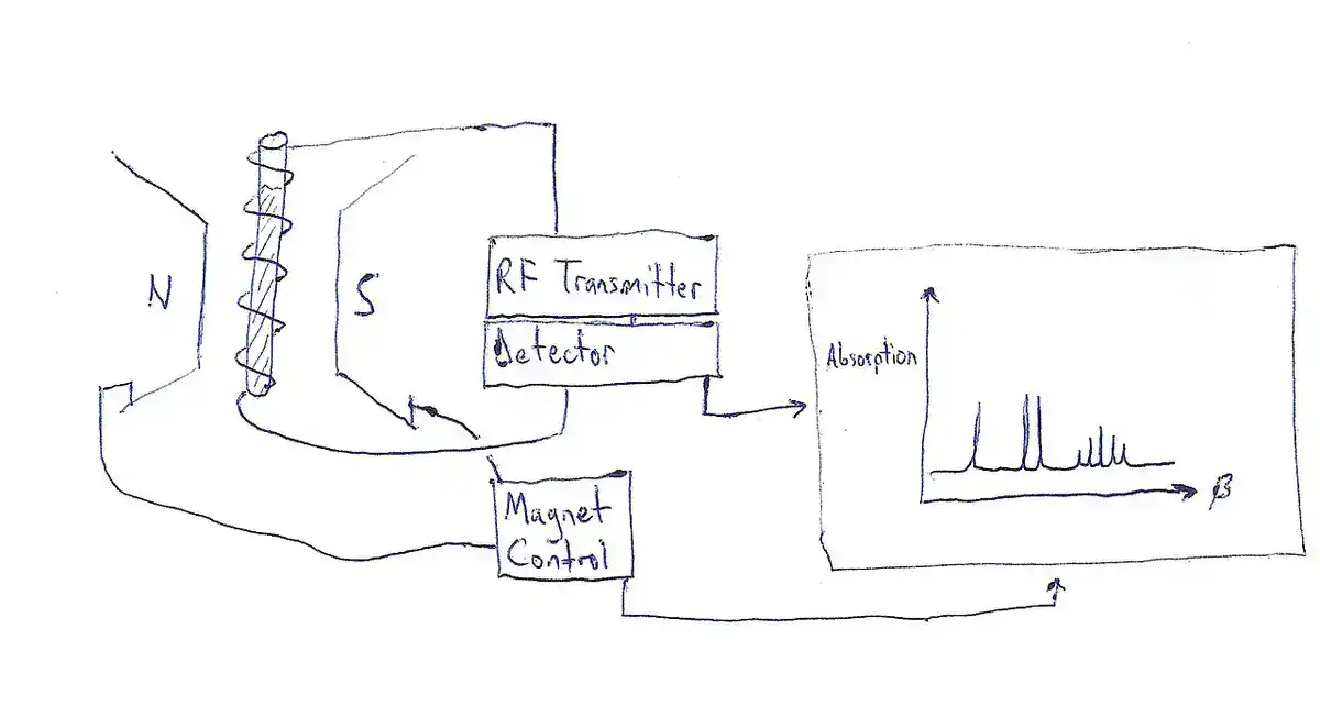
(CSIRO, CC BY 3.0
NMR spectroscopy is especially useful for studying molecules in solution, including dynamics and binding changes. An NMR protocol should state isotope labeling if used, buffer, concentration, temperature, pulse sequence, assignment method, controls, and whether the result is structural, dynamic, or binding-focused.
Cryo-electron microscopy, or cryo-EM, is useful for large complexes, multiple conformational states, membrane proteins, viruses, and particles that are difficult to crystallize. A cryo-EM protocol should state sample preparation, grid type, vitrification conditions, microscope settings, dose, particle picking, classification, reconstruction, map sharpening, resolution estimate, and model validation.
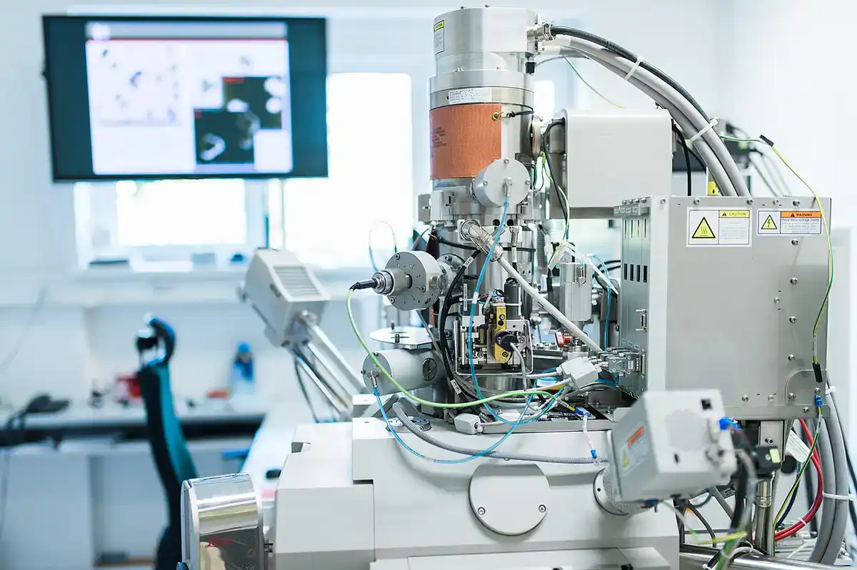
Structural methods should not be chosen only by prestige. X-ray crystallography, NMR, and cryo-EM answer overlapping but different questions. A stable small soluble protein, a flexible protein complex, a membrane protein, and a heterogeneous assembly may each need a different strategy.
Small-Angle Scattering and Integrative Structural Methods
Small-angle X-ray scattering, often called SAXS, measures how X-rays scatter from macromolecules in solution. It gives low-resolution information about size, shape, flexibility, and oligomeric state. SAXS is often used alongside crystallography, NMR, cryo-EM, SEC-MALS, mass spectrometry, crosslinking, or computational modeling.
A useful SAXS protocol should state sample purity, concentration series, buffer match, radiation-damage check, aggregation check, subtraction method, radius of gyration analysis, pair-distance distribution, model fitting, and whether the sample is monodisperse enough for interpretation.
SAXS is powerful but easy to overclaim. It rarely gives atomic structure by itself. Its strength is in solution-state shape, flexibility, and integrative modeling when paired with other evidence.
Circular Dichroism and Protein Folding Protocols
Circular dichroism spectroscopy, or CD, is used to study protein secondary structure, folding, conformational change, and thermal stability. Far-UV CD is commonly used to estimate alpha helix, beta sheet, and random coil content. Near-UV CD can provide information about tertiary structure around aromatic residues and disulfide bonds.
A useful CD protocol should state protein concentration, buffer absorbance, cuvette path length, wavelength range, scan speed, temperature, baseline correction, number of scans, smoothing rule, and whether the experiment measures structure, stability, ligand effect, or folding transition.
Common CD mistakes include using absorbing buffers, measuring too concentrated a sample, ignoring high-tension voltage limits, over-smoothing spectra, comparing spectra without baseline correction, and treating secondary-structure estimates as exact structural models.
Fluorescence Spectroscopy and FRET Protocols
Fluorescence spectroscopy measures light emitted by fluorophores after excitation. In biophysics, fluorescence can report on protein folding, ligand binding, membrane environment, ion concentration, molecular motion, conformational change, and interaction between molecules.
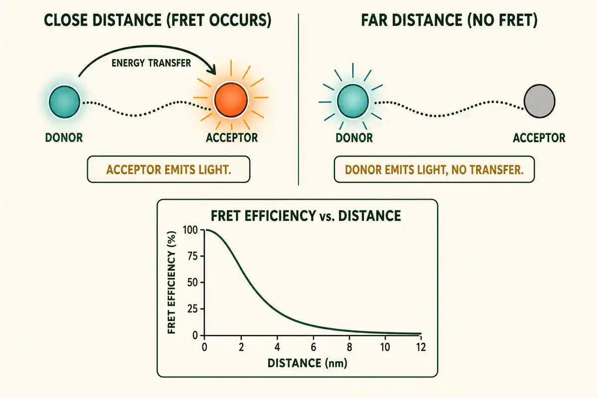
FRET, or Förster resonance energy transfer, is used as a distance-sensitive method when a donor fluorophore transfers energy to an acceptor fluorophore nearby. It is often used to study protein-protein interactions, nucleic acid folding, conformational changes, receptor activation, and molecular machines.
A useful fluorescence or FRET protocol should state excitation and emission wavelengths, fluorophore identity, labeling strategy, labeling efficiency, donor-only control, acceptor-only control, bleed-through correction, concentration, buffer, temperature, photobleaching check, inner-filter correction if relevant, and analysis model.
Fluorescence changes do not automatically prove binding or distance change. Changes can come from photobleaching, pH, quenching, aggregation, concentration effects, buffer changes, temperature, background, or spectral overlap. Controls decide whether the interpretation is believable.
Surface Plasmon Resonance and Binding Kinetics
Surface plasmon resonance, or SPR, is a label-free optical method used to measure molecular interactions in real time. Harvard's Center for Macromolecular Interactions describes SPR as an optical technique for measuring molecular interactions and notes that it can measure kinetic binding constants and equilibrium binding constants. See Harvard CMI Surface Plasmon Resonance.
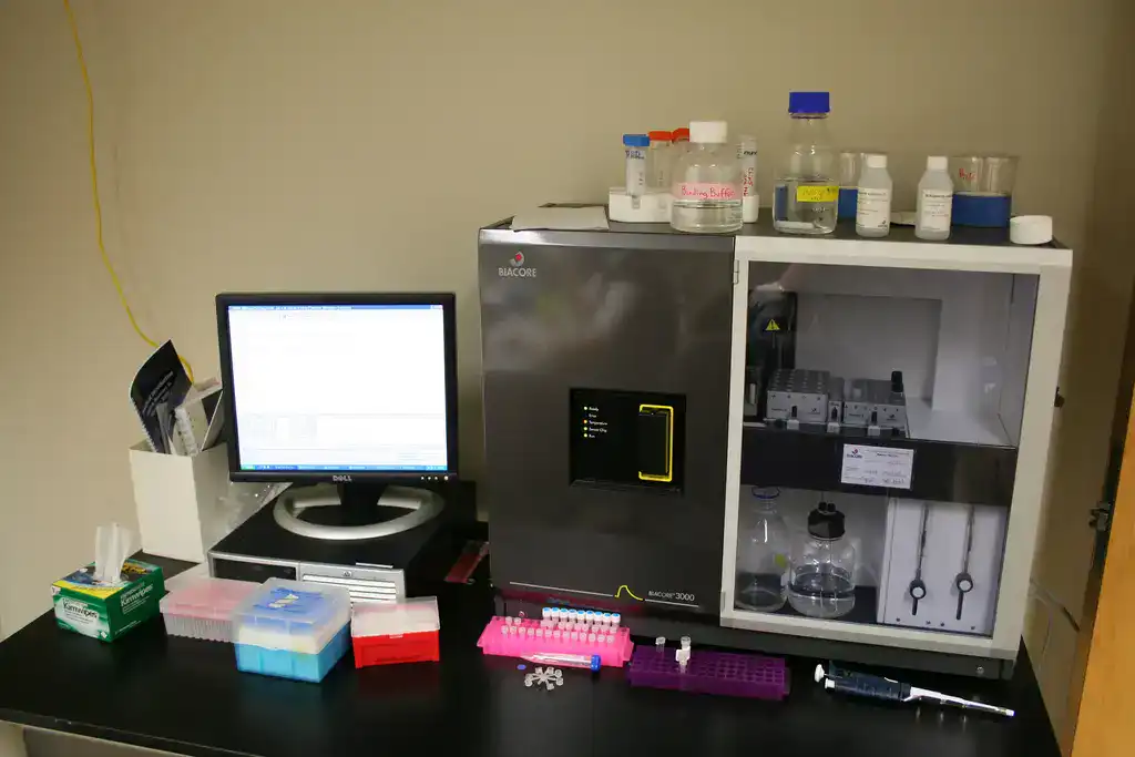
SPR is commonly used for protein-protein interactions, antibody-antigen binding, receptor-ligand binding, protein-DNA interactions, small-molecule binding, epitope binning, and drug-discovery screening. It can report association rate, dissociation rate, affinity, specificity, concentration, and binding behavior across time.
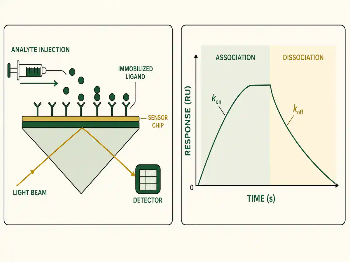
A useful SPR protocol should state ligand, analyte, immobilization chemistry, sensor surface, reference channel, buffer, flow rate, temperature, concentration series, injection time, dissociation time, regeneration condition, blank subtraction, reference subtraction, kinetic model, mass-transport check, and quality criteria.
SPR data can look convincing while being wrong. Common problems include nonspecific binding, poor reference surface, ligand overloading, mass-transport limitation, rebinding, buffer mismatch, analyte aggregation, poor regeneration, and forcing a 1:1 binding model onto a more complex interaction.
Isothermal Titration Calorimetry and Binding Thermodynamics
Isothermal titration calorimetry, or ITC, measures heat released or absorbed during molecular interactions. Malvern Panalytical describes ITC as a label-free technique that can determine binding affinity, stoichiometry, enthalpy, and entropy in solution. See Isothermal Titration Calorimetry.
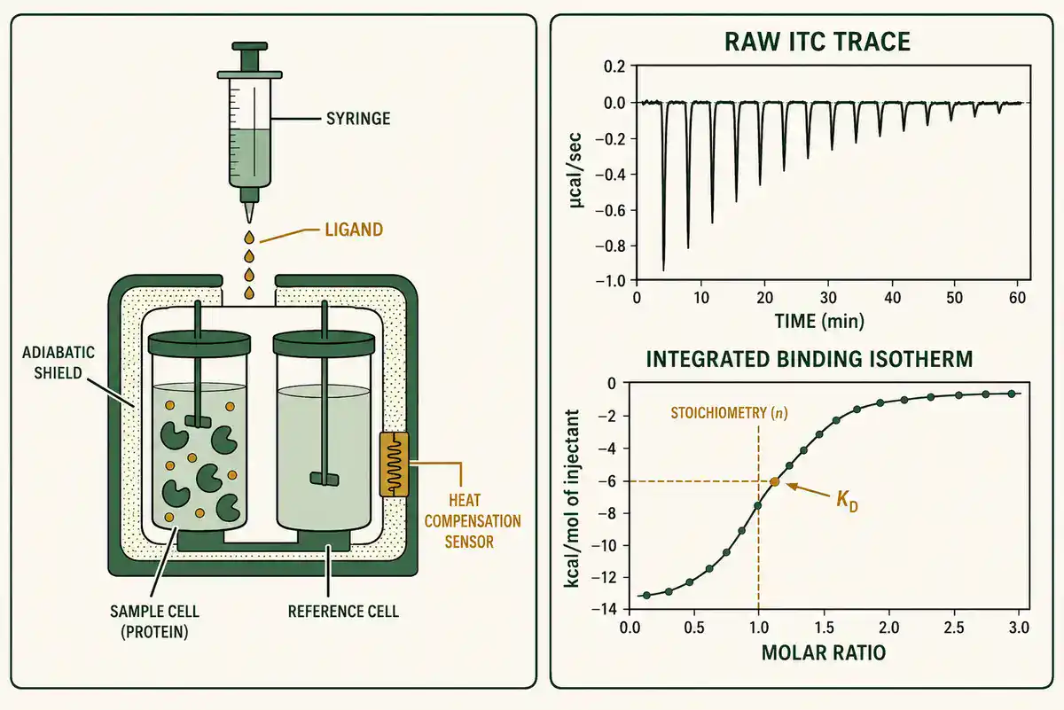
ITC is useful because it measures binding thermodynamics without requiring fluorescent or immobilized labels. It is often used for protein-ligand, protein-protein, protein-DNA, protein-RNA, lipid, carbohydrate, drug, and inhibitor interactions.
A useful ITC protocol should state sample purity, concentration accuracy, buffer matching, degassing if used, cell and syringe contents, injection volume, spacing, temperature, stirring speed, blank titration, heat-of-dilution correction, fitting model, replicate strategy, and expected binding window.
Common ITC mistakes include wrong concentration, poor buffer matching, weak heat signal, aggregation during titration, ligand insolubility, inaccurate baseline subtraction, using a model that does not match the interaction, and overinterpreting poorly constrained fits.
Differential Scanning Calorimetry and Thermal Stability
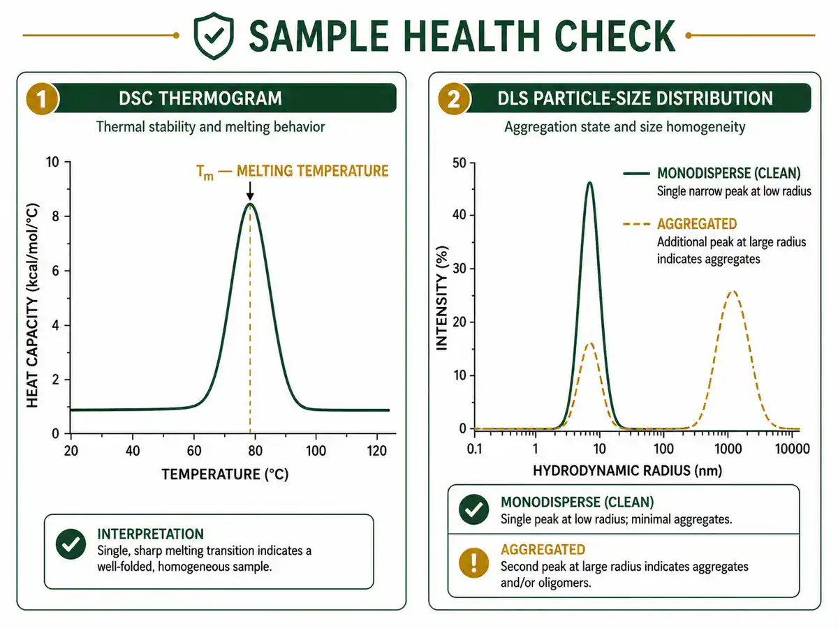
Differential scanning calorimetry, or DSC, measures heat capacity changes as a sample is heated or cooled. In biophysics, DSC is used to study protein unfolding, thermal transitions, stability, ligand effects, membrane phase transitions, and formulation behavior.
A useful DSC protocol should state sample concentration, buffer, scan rate, temperature range, baseline scan, reversibility check, replicate plan, transition temperature, enthalpy estimate, and whether unfolding is reversible or irreversible.
DSC can reveal stability changes, but it does not automatically explain why stability changed. Pairing DSC with CD, fluorescence, activity assays, mass spectrometry, or structural methods can make the interpretation stronger.
Dynamic Light Scattering and SEC-MALS
Dynamic light scattering, or DLS, estimates particle size in solution by measuring fluctuations in scattered light caused by Brownian motion. It is commonly used to detect aggregation, check sample monodispersity, compare buffer conditions, and screen formulations before structural or binding experiments.
SEC-MALS, or size-exclusion chromatography with multi-angle light scattering, is used to estimate molecular weight and oligomeric state in solution. It is valuable for distinguishing monomer, dimer, oligomer, aggregation, and complex formation.
A useful DLS or SEC-MALS protocol should state sample concentration, filtration or centrifugation, buffer, temperature, measurement angle or detector setup, chromatography column if used, calibration or normalization, baseline handling, and aggregation criteria.
DLS is sensitive to a small amount of large particles. A strong aggregation signal can dominate the result even when most molecules are smaller. That makes DLS excellent for detecting sample problems, but risky as the only method for defining a complex mixture.
Atomic Force Microscopy and Force Spectroscopy
Atomic force microscopy, or AFM, uses a sharp probe to image surfaces and measure nanoscale mechanical properties. In biology, AFM can be used to study cell surfaces, membranes, proteins, DNA, extracellular matrix, biomaterials, bacterial surfaces, cell stiffness, adhesion, and force-distance behavior.
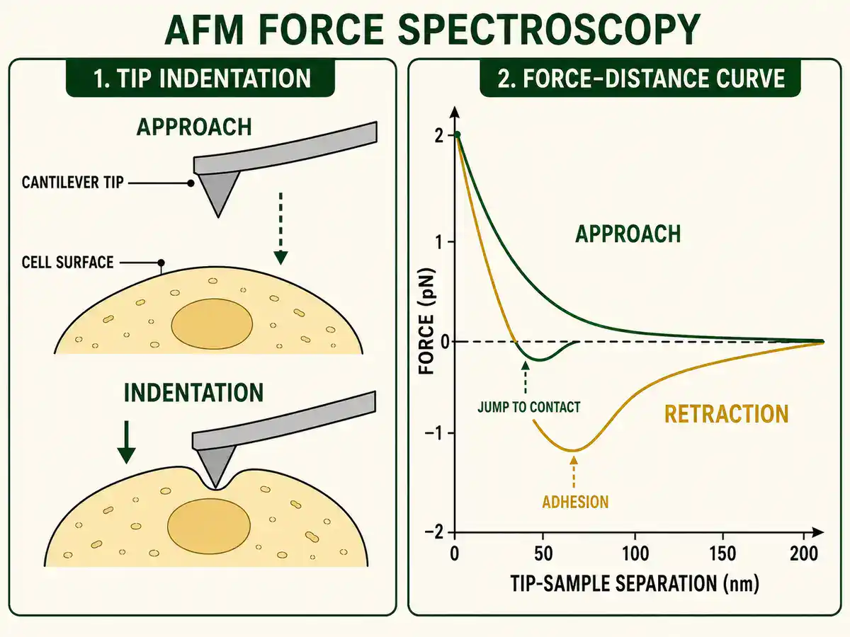
Bruker describes BioAFM platforms as tools for high-resolution imaging and nanomechanical analysis of biological samples, and notes that AFM can be integrated with optical techniques such as confocal and super-resolution microscopy. See Bruker BioAFM CellWizard Stage.
A useful AFM protocol should state sample immobilization, probe type, spring constant calibration, imaging mode, force setpoint, scan rate, buffer, temperature, indentation model, contact point selection, replicate strategy, and surface-damage check.
AFM is powerful because it can measure physical properties directly, but it is also sensitive to sample preparation, probe choice, surface attachment, force applied, drift, hydration, and model assumptions. For soft biological samples, the measurement itself can disturb the sample.
Optical Tweezers and Single-Molecule Force Methods
Optical tweezers use focused laser light to trap and manipulate microscopic particles. In biophysics, they can measure forces involved in DNA stretching, molecular motors, protein unfolding, ligand binding, and mechanical properties of biological polymers.
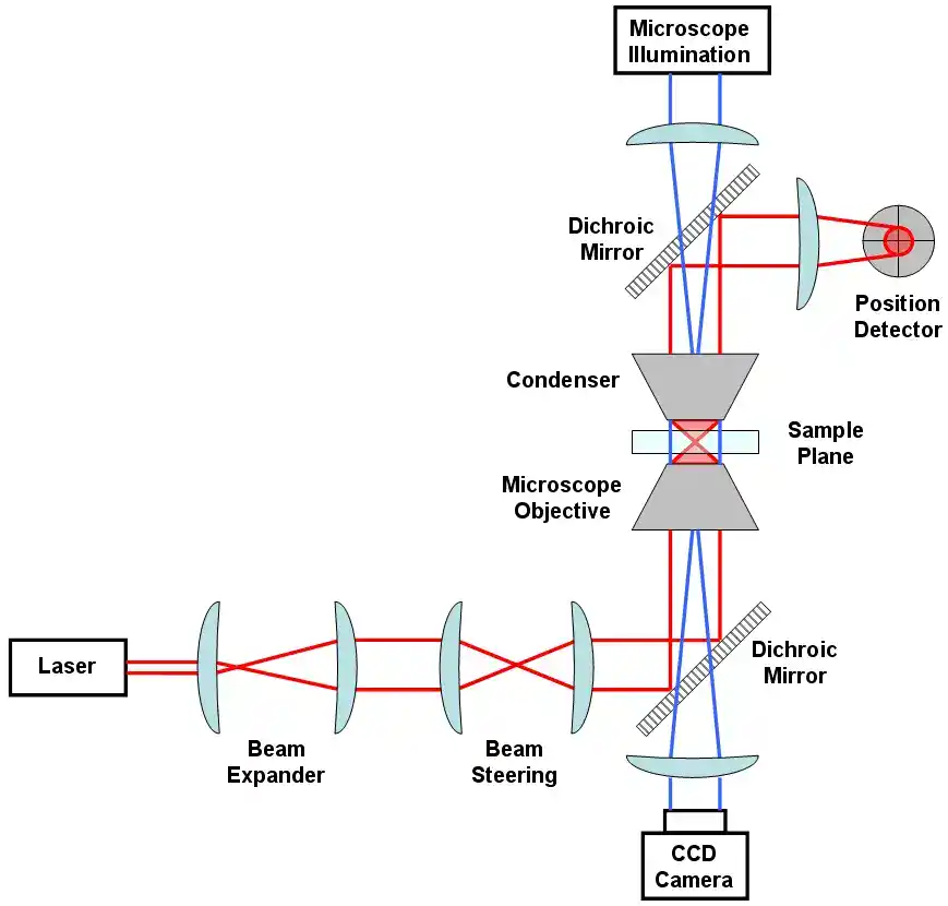
Single-molecule methods are useful because they can reveal heterogeneity that is hidden in bulk averages. A population-level measurement may show one average behavior, while single-molecule data reveal subpopulations, pauses, transitions, rare states, or force-dependent behavior.
A useful optical tweezers protocol should state bead chemistry, tether design, laser power, trap stiffness calibration, force calibration, sample chamber design, buffer, drift correction, pulling speed, data filtering, and rupture-force or extension analysis.
Single-molecule force data should be interpreted carefully. Attachment geometry, nonspecific binding, bead handling, heating, photodamage, pulling rate, and calibration choices can strongly affect the result.
Electrophysiology and Membrane Biophysics
Electrophysiology protocols measure electrical activity in cells, membranes, and ion channels. Patch clamp, voltage clamp, current clamp, extracellular recordings, planar lipid bilayer recordings, and membrane-potential assays are used in neuroscience, cardiac biology, pharmacology, membrane biology, sensory biology, and ion-channel research.
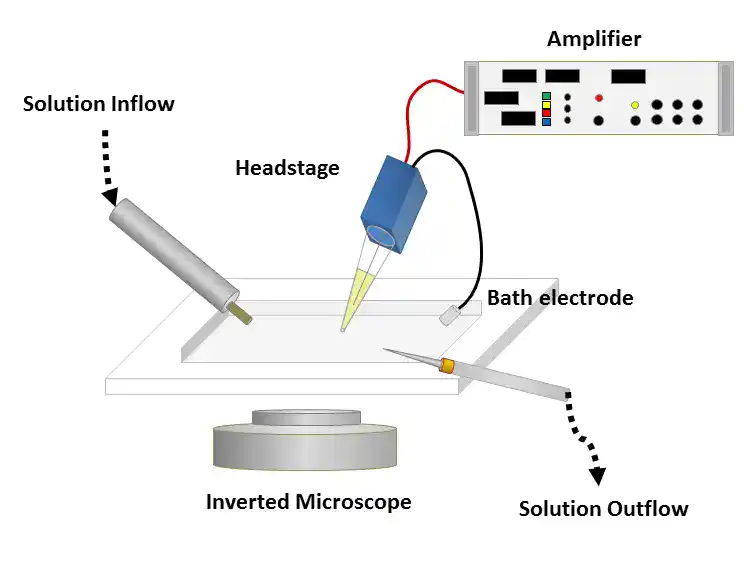
A useful patch clamp protocol should state cell type, recording configuration, electrode resistance, internal and external solutions, holding potential, voltage protocol, leak subtraction, series resistance, capacitance compensation, temperature, drug application method, inclusion criteria, and analysis model.
Membrane biophysics protocols may also measure ion transport, membrane permeability, lipid phase behavior, vesicle fusion, membrane protein activity, and membrane mechanics. The protocol should make clear whether the readout is electrical, optical, biochemical, mechanical, or computational.
Electrophysiology is highly quantitative, but fragile. Small changes in solution composition, temperature, seal quality, series resistance, cell health, or leak current can change the interpretation.
Molecular Dynamics and Computational Biophysics
Molecular dynamics simulations use physics-based models to simulate how atoms and molecules move over time. Computational biophysics can study protein folding, ligand binding, membrane proteins, nucleic acids, conformational change, ion channels, allostery, and molecular recognition.
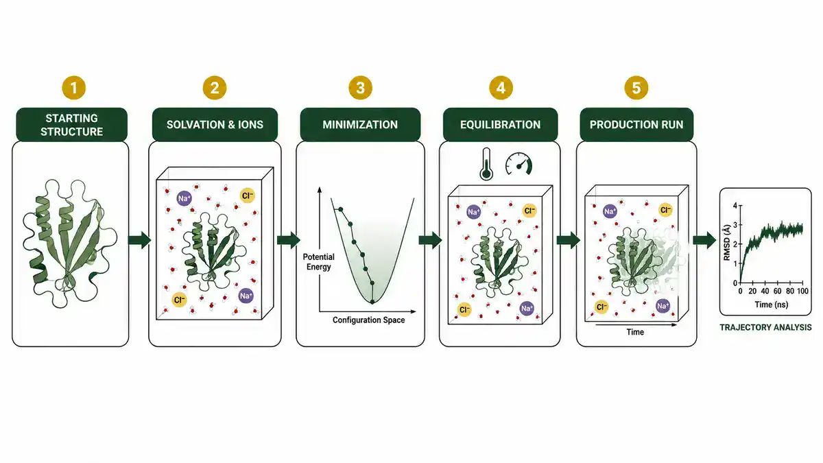
A useful molecular dynamics protocol should state the starting structure, force field, protonation states, solvent model, ion conditions, box size, minimization, equilibration, production run length, temperature and pressure control, constraints, replicates, convergence checks, and analysis metrics.
Common analysis outputs include RMSD, RMSF, radius of gyration, hydrogen bonds, distances, angles, solvent exposure, clustering, principal components, free-energy estimates, and interaction maps.
Simulations are not experiments, and they are not guesses either. They are model-based calculations. Their value depends on the quality of the starting structure, force field, sampling, assumptions, and agreement with experimental evidence.
How to Choose the Right Biophysics Protocol
Do not choose a biophysics method only because it looks advanced. Choose it because it matches the biological question, sample, physical parameter, concentration range, instrument access, and model assumptions.
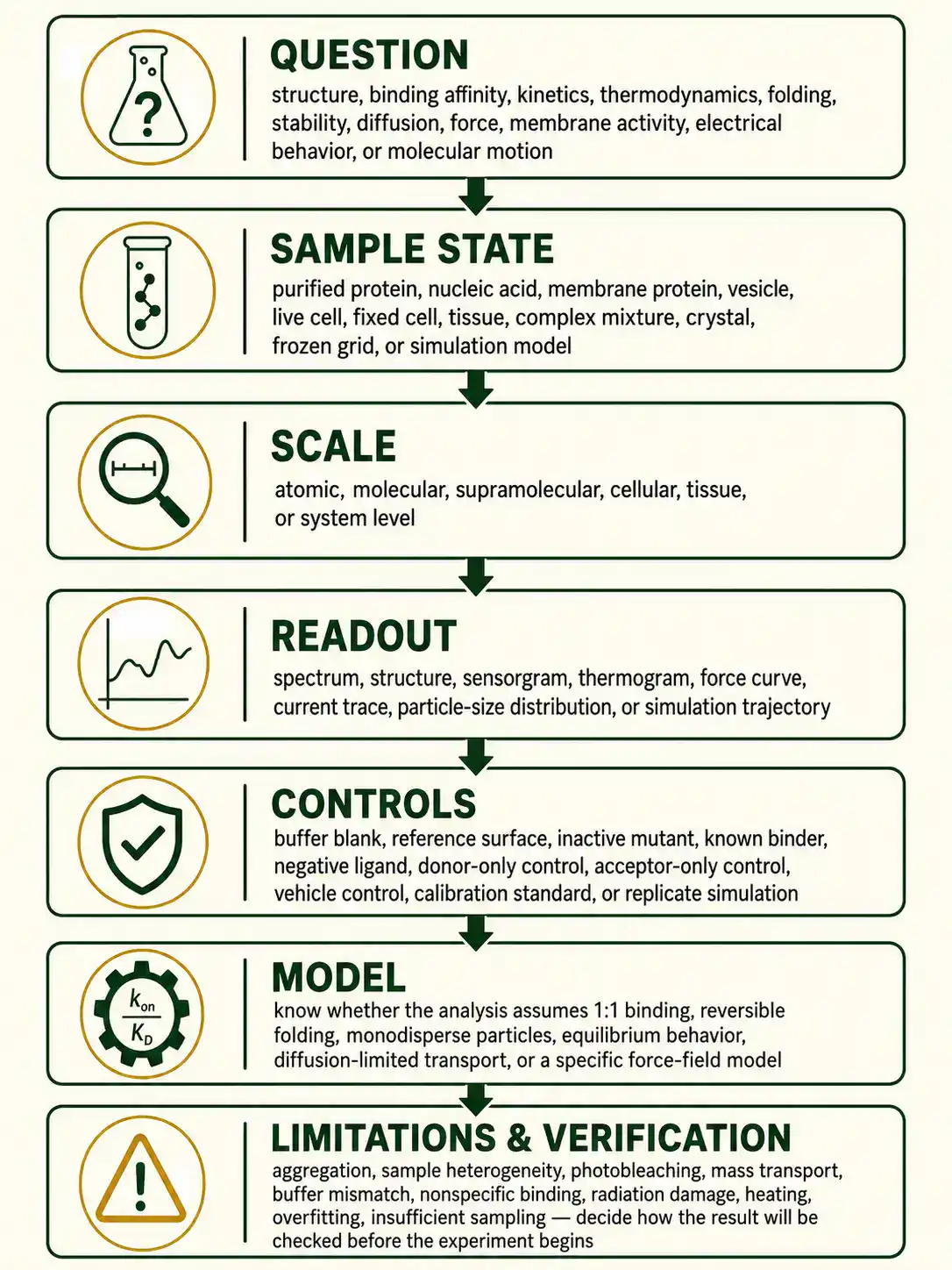
Before using any biophysics protocol, check these points:
- Question: structure, binding affinity, kinetics, thermodynamics, folding, stability, diffusion, force, membrane activity, electrical behavior, or molecular motion.
- Sample state: purified protein, nucleic acid, membrane protein, vesicle, live cell, fixed cell, tissue, complex mixture, crystal, frozen grid, or simulation model.
- Scale: atomic, molecular, supramolecular, cellular, tissue, or system level.
- Readout: spectrum, structure, sensorgram, thermogram, force curve, current trace, particle-size distribution, or simulation trajectory.
- Controls: buffer blank, reference surface, inactive mutant, known binder, negative ligand, donor-only control, acceptor-only control, vehicle control, calibration standard, or replicate simulation.
- Model: know whether the analysis assumes 1:1 binding, reversible folding, monodisperse particles, equilibrium behavior, diffusion-limited transport, or a specific force-field model.
- Limitations: aggregation, sample heterogeneity, photobleaching, mass transport, buffer mismatch, nonspecific binding, radiation damage, heating, overfitting, or insufficient sampling.
- Verification: decide how the result will be checked before the experiment begins.
Common Mistakes in Biophysics Protocols
Biophysical methods often produce numerical results, which can make weak data look more certain than it is. The numbers are only as good as the sample, controls, calibration, and model.
- Ignoring sample heterogeneity: aggregation, degradation, oligomer mixtures, or conformational mixtures can distort results.
- Using the wrong concentration range: binding and stability experiments need concentrations that match the method and expected KD.
- Skipping buffer matching: mismatched buffers can create false heat signals, refractive-index shifts, baseline changes, or fluorescence artifacts.
- Overfitting binding data: a complex model can fit noise if controls and replicates are weak.
- Assuming a 1:1 model: many biological interactions involve cooperativity, multiple sites, oligomerization, or conformational change.
- Trusting one method alone: a strong biophysical claim is often supported by orthogonal methods.
- Confusing affinity with kinetics: two interactions can have similar KD values but very different association and dissociation rates.
- Ignoring instrument calibration: force, light intensity, temperature, wavelength, detector response, and concentration calibration all matter.
- Forgetting negative controls: nonspecific binding and background can look like real signal.
- Overinterpreting structural models: resolution, flexibility, missing density, disorder, and model validation must be considered.
- Using simulations without validation: molecular dynamics results should be checked against experimental or independent computational evidence when possible.
- Publishing incomplete methods: missing buffer, concentration, temperature, model, software, or instrument settings makes biophysical data hard to reproduce.
Biophysics Calculators and Lab Tools
Biophysics protocols often depend on calculations. A wrong concentration, binding model, kinetic constant, force conversion, diffusion estimate, FRET distance, or electrophysiology equation can change the interpretation of the experiment.
Useful biophysics calculators include:
- binding affinity KD calculator
- association and dissociation rate calculator
- SPR kinetics calculator
- Hill equation calculator
- Scatchard plot calculator
- ITC thermodynamics calculator
- Gibbs free energy calculator for binding
- protein-ligand stoichiometry calculator
- FRET efficiency calculator
- FRET distance calculator
- fluorescence anisotropy calculator
- fluorescence quenching Stern-Volmer calculator
- circular dichroism mean residue ellipticity calculator
- protein melting temperature comparison tool
- DLS hydrodynamic radius calculator
- Stokes-Einstein diffusion calculator
- Brownian motion displacement calculator
- SEC-MALS molecular weight helper
- sedimentation coefficient calculator
- Nernst equation calculator
- Goldman-Hodgkin-Katz voltage calculator
- membrane potential calculator
- patch clamp conductance calculator
- ion flux calculator
- osmotic pressure calculator
- optical trap stiffness calculator
- force-extension curve calculator
- AFM indentation modulus calculator
- molecular dynamics RMSD calculator
- molecular dynamics RMSF calculator
- radius of gyration calculator
- protein concentration and dilution calculator
- Beer-Lambert absorbance calculator
- buffer ionic strength calculator
- Debye length calculator
BioExplorer's Biology Tools and Calculators hub is building free browser-based tools by branch of biology. As BioExplorer expands its biophysics tools, this page can link directly to binding, FRET, SPR, ITC, diffusion, membrane-potential, electrophysiology, and molecular-simulation calculators.
Trusted Biophysics Protocol Resources
Use BioExplorer as a guide, but always check your institution's approved SOPs, instrument manuals, core-facility guidance, software documentation, safety rules, and supervisor instructions before performing real laboratory or computational work. These external resources are useful starting points for biophysics methods and protocol background:
- Biophysical Society: What Is Biophysics?: overview of biophysics as a field connecting physics, chemistry, mathematics, engineering, and biology.
- Nature Protocols: peer-reviewed protocol articles with design advice, troubleshooting, data analysis, limitations, and result interpretation.
- PDB-101 Methods for Determining Structure: educational guide to X-ray crystallography, NMR spectroscopy, electron microscopy, and related structural methods.
- Malvern Panalytical ITC Guide: overview of isothermal titration calorimetry for label-free binding thermodynamics.
- Harvard CMI Surface Plasmon Resonance: explanation of SPR for real-time interaction analysis and kinetic binding constants.
- Bruker BioAFM: overview of AFM tools for high-resolution biological imaging and nanomechanical analysis.
- RCSB Protein Data Bank: public archive of experimentally determined 3D structures of biological macromolecules.
- Biology Methods and Protocols: Oxford Academic journal covering experimental methods across biological research.
Safety and Responsibility
Biophysics protocols can involve lasers, UV light, X-rays, cryogens, high voltage, strong magnets, vacuum systems, pressurized gases, nanoparticles, toxic stains, biological samples, recombinant proteins, living cells, membrane preparations, computational models, and large datasets. This page is educational. It does not replace formal training, institutional SOPs, instrument manuals, chemical safety guidance, biosafety approval, radiation safety training, laser safety training, or supervision by qualified personnel.
For real laboratory work, follow approved procedures for sample handling, instrument operation, biological material, chemical waste, laser use, cryogen handling, radiation safety, and data reporting. For computational work, document the model, software version, parameters, input files, and analysis scripts clearly enough for another person to understand the result.
Related BioExplorer Resources
- Biology Methods and Protocols: main hub for biology lab methods, protocol categories, safety notes, and trusted resources.
- Biochemistry Methods and Protocols: methods for protein assays, enzyme kinetics, SDS-PAGE, western blotting, chromatography, and protein purification.
- Molecular Biology Methods and Protocols: methods for DNA, RNA, PCR, gel electrophoresis, cloning, sequencing prep, and molecular analysis.
- Cell Biology Methods and Protocols: methods for cell culture, microscopy, flow cytometry, immunofluorescence, cell counting, and cell-based assays.
- Microscopy Methods and Protocols: guide to brightfield, phase contrast, fluorescence, confocal, electron microscopy, and sample preparation.
- Biology Tools and Calculators: free browser-based biology calculators and educational tools.
- Biochemistry Glossary: clear definitions of proteins, enzymes, amino acids, ATP, chromatography, and related biochemical terms.
- Molecular Biology Glossary: definitions of DNA, RNA, PCR, sequencing, genes, expression, and molecular analysis terms.
Frequently Asked Questions
Biophysics protocols are written workflows for studying biological systems with physical and quantitative methods, including structure determination, binding analysis, spectroscopy, membrane measurements, force measurements, electrophysiology, and molecular simulations.
Common biophysics methods include X-ray crystallography, NMR spectroscopy, cryo-EM, SAXS, circular dichroism, fluorescence spectroscopy, FRET, SPR, ITC, DSC, DLS, AFM, optical tweezers, patch clamp, and molecular dynamics simulations.
Biochemistry focuses on the chemical reactions and molecules of life. Biophysics focuses on the physical principles, forces, structures, dynamics, thermodynamics, and quantitative measurements that explain how biological molecules and systems behave.
Surface plasmon resonance is used to measure molecular binding in real time. It can estimate association rate, dissociation rate, binding affinity, specificity, and concentration when the experiment is properly controlled and modeled.
Isothermal titration calorimetry is used to measure heat released or absorbed during binding events. It can estimate binding affinity, stoichiometry, enthalpy, and entropy without requiring a fluorescent or immobilized label.
FRET is used to study short-range molecular distances, conformational changes, and interactions between molecules labeled with compatible donor and acceptor fluorophores.
X-ray crystallography usually requires ordered crystals and can produce high-resolution structures. NMR studies molecules in solution and can measure dynamics. Cryo-EM images frozen particles and is especially useful for large complexes, heterogeneous assemblies, and membrane proteins.
Controls help distinguish real physical or biological signal from aggregation, nonspecific binding, buffer mismatch, photobleaching, instrument drift, mass transport, sample degradation, poor calibration, or incorrect model fitting.
Not always. Online protocols may not match your sample, instrument, safety requirements, calibration state, or analysis model. For real work, follow approved SOPs, instrument manuals, facility guidance, safety rules, and supervisor instruction.
Cite this page
BioExplorer. (2026, August 26). Biophysics Methods and Protocols. https://www.bioexplorer.net/methods_and_protocols/biophysics/