


Generally, in the big schema things of the human body, the skin often does not strike as an organ. However, the skin is composed of tissues and performs mission-critical functions in the body.
The skin and their accessory structures such as hair, glands, and nails make up the integumentary system, which provides the body with overall protection.
The skin contains multiple layers of cells and tissues, which are attached to underlying structures such as blood vessels and bones by connective tissues.
In this article, we will explore the different types of skin cells (cells of the Epidermis), the layers of skin cells and the functions of the skin cells.
Table of Contents
Cells of the Epidermis
There are 4 types of skin cells in humans namely Keratinocytes, Melanocytes, Langerhans cells, and Merkel cells.

1. Keratinocytes

A keratinocyte is the primary cell type (about 90%) within the epidermis which is the outermost layer of the skin. The alternate names for keratinocytes are basal cells since they are found in the basal layer (the stratum basale) of the skin.
![]()
2. Melanocytes

Melanocytes are second types of skin cells which produce melanin. Melanin is a dark pigment contained inside melanosomes that determines the skin color of the person. They are found in the bottom layer of the skin’s epidermis.
The examples include the middle layer of the eye, the inner ear, meninges, bones, and the heart. Melanin protects against the Ultra-Violet (UV) radiation and support in the immune system.
![]()
3. Langerhans Cells
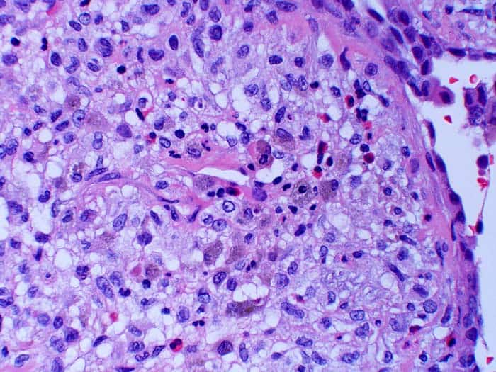
Langerhans cells are the third type of skin cells also known as dendritic cells (antigen-producing immune cells – APC). These cells contain organelles called Birbeck granules.
Langerhans cells are found in all layers of the epidermis. However, they are most prominent in the stratum spinosum layer (a layer between the stratum granulosum and stratum basale). Examples include blood vessels, the mucosa of the mouth, foreskin, and vaginal epithelium.
These Langerhans cells can also be found in other tissues, such as lymph nodes. Note that while similar in appearance, these langerhans cells’ structures different from the dendrites of neurons.
![]()
4. Merkel cells

Merkel cells are also called Merkel-Ranvier cells or tactile epithelial cells. They are oval-shaped cells responsible for light, touch, and sensation and found in the skin of vertebrates.
Merkel cells are in plentiful in highly sensitive skins and make synaptic contacts with somatosensory afferent nerve fibers. Although it is very uncommon, these cells may become malignant and form a Merkel cell carcinoma-an aggressive and difficult to treat skin cancer.
Though it has been found that Merkel cells are derived from neural crest cells, the more recent experiments in mammals have revealed that they are in fact from epithelial in origin.
![]()
Layers of the Skin
The skin is composed of 3 layers namely:

1. Epidermis
This is the outer most superficial layer which is made up of 5 inner layers. They are Stratum basale, Stratum spinosum, Stratum granulosum, Stratum lucidum, and Stratum corneum.
In general, skin’s epidermal layer is subjected to constant wear & tear from external factors such as sunlight, chemicals such as soaps, and pollution.
![]()
2. Dermis
Dermis cover the significant portion of the skin’s layer. The dermis layer has connective tissues, blood vessels, oil and sweat glands, nerves, hair follicles, and other structures.
The dermis is made up of two inner layers namely – a thin upper layer called the papillary dermis, and a thick lower layer called the reticular dermis.
![]()
3. Subcutaneous Layer
Subcutaneous layer is also known as hypodermis. The hypodermis is the innermost layer of the skin. This layer hosts fat and connective tissues that house larger blood vessels and various nerves.
The primary function of the hypodermis is to act as an insulator for regulating the body temperature.
![]()
Why are tattoos permanent though skin cells die and get replaced?
The answer lies hidden in the second layer of the skin (Dermis). The permanent tattoo ink is injected till the dermis layer so that it stays permanent. If it is put on the outer layer, then it will be worn out as time progresses. That is why permanent tattooing is always a harrowing and painful experience.
![]()
Functions of Skin Cells
Here are the main functions of the skin cells:
- Protection: The single most important function of the skin is the protection from the outside environment.
- Keeping Cool: The skin secretes the sweat when the body is hot thus keeping the coolness within by evaporating the sweat from the skin.
- Communication: The skin connects to the nerve fibers to transmit the sensation to/from the brain. (Example: touching a hot or cold surface)
- Reservoir of Nutrients: The skin holds the important fluids and nutrients to keep the skin healthy and functional (Example: Collagen and Elastin).
![]()
Cite This Page
References
- “Anatomy and Organization of Human Skin” by J.A.McGrath, R.A.J.Eady, F.M.Pope. Accessed October 23, 2018. Link.
- “5.1 Layers of the Skin | Anatomy & Physiology”. Accessed October 23, 2018. Link.
- “The origin of epidermal melanocytes. Implications for the histogenesis of nevi and melanomas. – PubMed – NCBI”. Accessed October 23, 2018. Link.
- “An improved ontological representation of dendritic cells as a paradigm for all cell types”. Accessed October 23, 2018. Link.
- “Pathophysiology of merkel cell Munde PB, Khandekar SP, Dive AA, Sharma A – J Oral Maxillofac Pathol”. Accessed October 23, 2018. Link.


