
Cellular Respiration Equation: Stages, Products, and ATP Yield
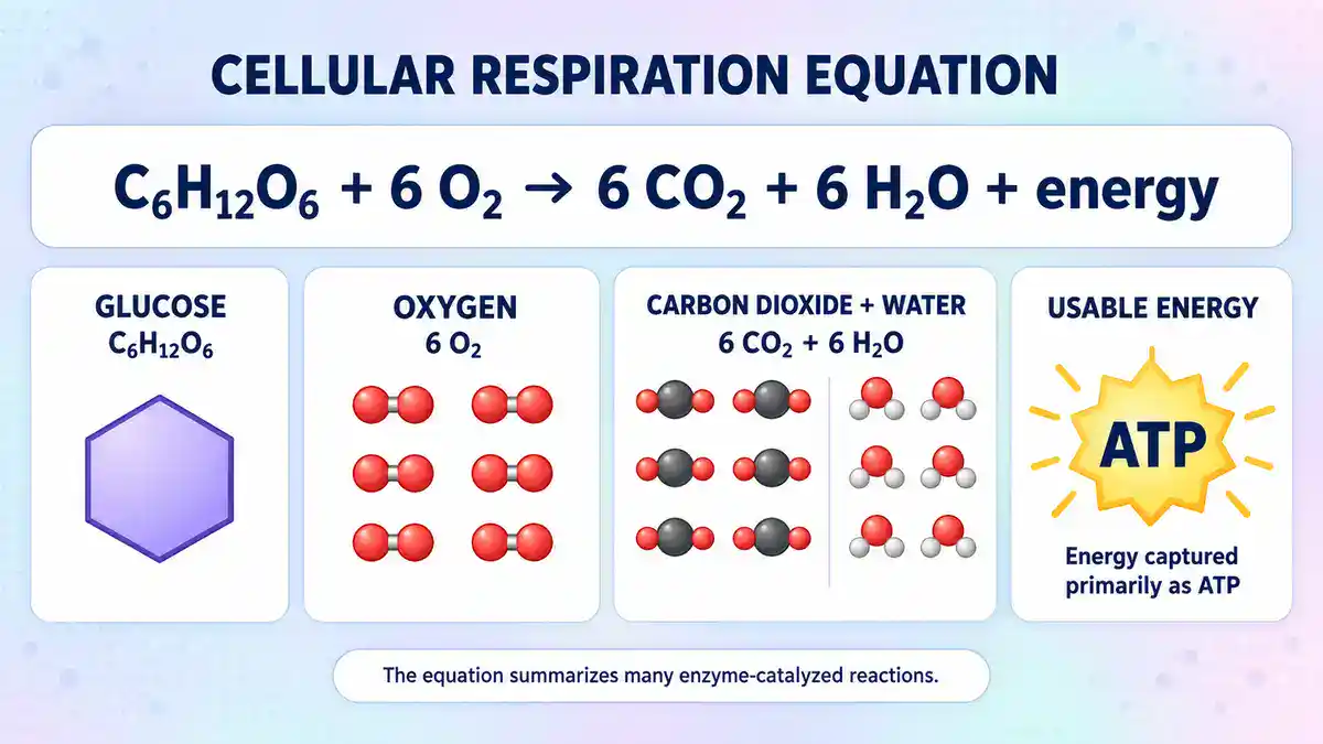
The cellular respiration equation is C6H12O6 + 6 O2 → 6 CO2 + 6 H2O + energy. Glucose and oxygen go in. Carbon dioxide and water come out. The cell keeps the energy as adenosine triphosphate, or ATP. In a typical mammalian cell, one glucose molecule yields an idealized 30 to 32 ATP. That figure is a convention, not a universal constant, and the reasons appear further down this page.
ATP is not a battery that stores energy in a bond. It is a carrier with a high phosphoryl-transfer potential. Transferring its terminal phosphate group to another molecule is thermodynamically favorable, because the products are more stable overall. Cells cannot stockpile it. A human body hydrolyzes an estimated 100 to 150 moles of ATP every day.[1] That amount dwarfs anything a cell could hold, so ATP must be regenerated continuously.
Cellular respiration is the controlled, stepwise oxidation of a fuel molecule, and it is what makes that regeneration possible. It is not a single chemical reaction. It is a set of linked pathways that pass electrons from glucose along a series of carriers to a final acceptor.
The energy released along the way drives ADP + Pi → ATP. Complete oxidation of one glucose molecule produces about 30 ATP. Close to half of the energy theoretically available from glucose is captured that way, and the rest is released as heat.[2]
One number here will look wrong to anyone taught from an older textbook. The figure of 36 or 38 ATP per glucose still appears in many classrooms. It has been superseded. The section on ATP yield explains where 38 came from and what replaced it.
Cellular Respiration Equation Guide:
- Summary of key facts
- What the cellular respiration equation actually says
- Why the "38 ATP" figure is out of date
- The four stages of cellular respiration
- Chemiosmosis: how a proton gradient becomes ATP
- Where each stage takes place
- Why the total is 30 in some cells and 32 in others
- What oxygen actually does
- Fermentation: keeping glycolysis running without oxygen
- Anaerobic respiration is a different process from fermentation
- Fuels other than glucose
- Cellular respiration and photosynthesis compared
- Why cellular respiration matters outside the classroom
- Common misconceptions
- Frequently asked questions
- Jump to comments
Summary of key facts
| Question | Answer |
|---|---|
| What is the cellular respiration equation? | C6H12O6 + 6 O2 → 6 CO2 + 6 H2O + energy |
| How much ATP does one glucose molecule yield? | An idealized 30 to 32 ATP in a typical mammalian cell, not 38 |
| What are the stages? | Glycolysis, pyruvate oxidation, the citric acid cycle, and the electron transport chain with oxidative phosphorylation |
| Where does each stage happen in a typical mammalian cell? | Glycolysis in the cytosol; the other three in the mitochondrion |
| Where does it happen in bacteria and archaea? | In many of them, glycolysis and the cycle in the cytoplasm, electron transport on the cytoplasmic membrane |
| What does oxygen do? | It accepts electrons at the end of the electron transport chain and is reduced to water |
| How much ATP does fermentation yield? | Two per glucose for homolactic and alcoholic fermentation through the Embden-Meyerhof-Parnas pathway. Other routes yield less or more |
| Is anaerobic respiration the same as fermentation? | No. Anaerobic respiration uses membrane-linked electron transfer to an acceptor other than oxygen. Fermentation uses none |
| Does lactic acid fermentation release CO2? | No. Strict homolactic fermentation of glucose produces lactate only |
| Do plants carry out cellular respiration? | Yes, continuously, in addition to photosynthesis |
What the cellular respiration equation actually says
The balanced equation, term by term
The standard summary equation for aerobic cellular respiration is:
C6H12O6 + 6 O2 → 6 CO2 + 6 H2O + energy
In words: one glucose molecule plus six oxygen molecules yield six carbon dioxide molecules, six water molecules, and usable energy.
The equation balances on atoms. The six carbons of glucose leave as six carbon dioxide molecules. The twelve hydrogens leave as hydrogen in six water molecules. Eighteen oxygen atoms sit on each side. Six come from glucose and twelve from the six O2 molecules.
Complete oxidation of glucose releases a large amount of free energy, with a standard free energy change near minus 686 kilocalories per mole. Cells oxidize glucose in a series of steps coupled to ATP synthesis, which is how they capture part of that energy.[3]
Why the equation is a summary and not a single reaction
No enzyme performs the reaction as written. Burning glucose in a flame does roughly the same chemistry in a single step, and all the energy escapes as heat and light. A cell instead removes electrons a few at a time, across dozens of enzyme-catalyzed reactions, so the energy arrives in packets small enough to trap in ATP.
Two details are worth pinning down early. First, the six water molecules are a net total from the atom balance. They are not a running count of each step. The electron transport chain alone forms twelve water molecules from six O2. Water is also consumed at several points in the citric acid cycle. No single stage matches the number in the equation.
Second, cell biology textbooks write the right side as “energy” rather than a number of ATP. Part of the reason is arithmetic. Each ADP and phosphate pair joined into ATP releases a water molecule. Writing ATP into the equation therefore raises the water count on the product side.
Glucose is the convention, not the rule
Glucose is written into the equation for two reasons: it is the reference fuel used to teach the pathway, and it is the sugar that circulates in blood. Cells oxidize fatty acids, amino acids, other sugars, and stored glycogen through the same core machinery. A later section covers what changes when the fuel is fat or protein.
Why the “38 ATP” figure is out of date
Where 36 and 38 came from
The older figure rests on two round numbers. One NADH was taken to yield three ATP. One FADH2 was taken to yield two. Breaking down glucose through glycolysis, pyruvate oxidation and the citric acid cycle produces four ATP directly, ten NADH, and two FAD-linked electron pairs.[4]
Ten NADH at three ATP gives 30. Two FADH2 at two ATP gives four. Adding the four direct ATP gives 38. Subtracting two for a costlier route into the mitochondrion gives 36. The arithmetic is tidy. That tidiness is part of why it survived so long.
What the measured numbers turned out to be
The values of 3 and 2 were not invented out of nothing. They were conventional interpretations of genuinely difficult early measurements, and the integer form belonged to the interpretation rather than to the data. Modern accounting starts from protons instead. Consensus values are 10 protons pumped per NADH oxidized and 6 per succinate.
The synthase itself uses about 3 protons per ATP. Importing ADP and phosphate while exporting ATP costs one more proton. That gives P/O ratios of 10 divided by 4, or 2.5 for NADH, and 6 divided by 4, or 1.5 for succinate. Both sit well below the older textbook values of 3 and 2. Careful re-measurement has since confirmed experimental values close to 2.5 and 1.5.[5]
Two consequences follow. The yield per glucose drops by several ATP. It also stops being a whole number, because the underlying ratios are not whole numbers.
The modern arithmetic, stage by stage
The table below uses 2.5 ATP per mitochondrial NADH and 1.5 per FAD-linked pair. It describes an idealized aerobic mammalian cell.
| Stage | ATP made directly | NADH | FAD-linked pairs | ATP from oxidative phosphorylation |
|---|---|---|---|---|
| Glycolysis (cytosol) | 2 | 2 (cytosolic) | 0 | 3 or 5, depending on the shuttle used |
| Pyruvate oxidation (matrix) | 0 | 2 | 0 | 5 |
| Citric acid cycle (matrix) | 2 | 6 | 2 | 18 |
| Total | 4 | 10 | 2 | 26 to 28 |
Four ATP made directly, plus 26 to 28 from oxidative phosphorylation, gives 30 to 32 ATP per glucose. Oxidative phosphorylation is the process that harnesses the reduction of oxygen to make ATP.
Approximately 30 to 32 ATP is the figure current references give, using about 2.5 ATP per NADH and about 1.5 per FADH2.[6] A later section explains why the range has two ends.
What careful modeling gives, and why real cells fall short
Detailed modeling puts the ceiling slightly higher than the textbook range. Complete oxidation of glucose by cells yields up to 33.45 ATP per glucose, at a maximum P/O of 2.79. Of that, 2 ATP come from Glycolysis and up to 31.45 from the oxidative reactions.[7]
That is a maximum under ideal coupling. It is not a routine output. Real mitochondrion leak protons back across the inner membrane. Some electrons take routes that bypass a proton pump.
The same correction applies outside animals. One balance-sheet model puts the potential yield of plant respiration near 27.5 ATP per hexose. The figure falls further when a quarter of oxygen uptake runs through the alternative oxidase. The author concludes that plant respiratory ATP yield is smaller than often assumed, and certainly less than older textbook values of 36 to 38 per hexose.[8]
So the defensible statement is a range with a reason attached. About 30 to 32 ATP per glucose is the standard modern convention for a mammalian cell. The ideal ceiling sits a little above it. What a real cell achieves at any moment is lower still, and it varies by tissue and coupling state.
The four stages of cellular respiration
Some summaries describe three stages. They fold the conversion of pyruvate to acetyl-CoA into either glycolysis or the citric acid cycle. Treating it as its own stage is clearer here. It happens in a different compartment from glycolysis and uses a different enzyme complex.
Stage 1: Glycolysis splits glucose into two pyruvate molecules
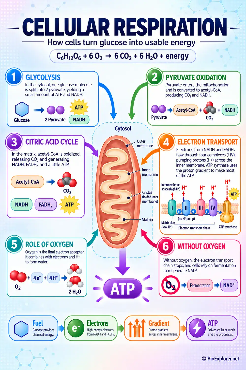
Glycolysis takes its name from the Greek roots for sweet and for loosening. It occurs in the cytosol and requires no oxygen. Two ATP are consumed in an early investment phase. Four are produced in a later payoff phase. The pathway yields 4 ATP, 2 NADH, and 2 pyruvates per glucose, for a net gain of two ATP. Three steps are effectively irreversible and act as control points. They are catalyzed by hexokinase, by phosphofructokinase-1, and by pyruvate kinase.[9]
Stated in one line, glycolysis converts one glucose molecule into two pyruvate molecules with a net production of two ATP.[10]
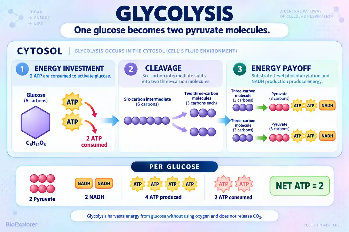 Glycolysis infographic showing one glucose molecule split into two pyruvate in the cytosol, with 2 ATP consumed, 4 ATP produced, 2 NADH formed, and a net gain of 2 ATP.
Phosphofructokinase-1 is often called the rate-limiting step. That is outdated shorthand. It is better described as a committed and heavily regulated step. Control of glycolytic flux is distributed rather than concentrated. One systematic study overexpressed each of the twelve enzymes linking external glucose to excreted lactate. It found substantial flux control at four steps: glucose import, hexokinase, phosphofructokinase, and lactate export. It found none at any step of lower glycolysis.[11]
Two features matter for everything that follows. The two ATP are made by substrate-level phosphorylation. A phosphate group moves directly from a substrate to ADP, with no membrane and no oxygen involved. Separately, the cytosol holds only a limited pool of NAD+. It must be reoxidized continuously or the pathway stalls. What happens to that NADH is the fork between respiration and fermentation.
Stage 2: Pyruvate oxidation links glycolysis to the cycle
Pyruvate is transported from the cytosol into the mitochondrial matrix. There the pyruvate dehydrogenase complex converts pyruvate to acetyl-CoA and CO2.[12] Each pyruvate loses one carbon as carbon dioxide. The remaining two-carbon acetyl group attaches to coenzyme A. One NAD+ is reduced to NADH.
Glycolysis produced two pyruvates, so this stage runs twice per glucose. It contributes 2 CO2 and 2 NADH. Those two NADH are easy to overlook. They come from neither glycolysis nor the cycle.
Stage 3: The citric acid cycle strips the remaining electrons
The citric acid cycle is also called the Krebs cycle or the tricarboxylic acid cycle. It accepts the acetyl group from acetyl-CoA and oxidizes it completely. The cycle has eight steps. Seven sit in the mitochondrial matrix. The outlier is succinate dehydrogenase, which is bound to the inner mitochondrial membrane and forms part of the electron transport chain.
Its main control points are the pyruvate dehydrogenase complex that feeds it, isocitrate dehydrogenase, and the alpha-ketoglutarate dehydrogenase complex.[13]
That location matters, but it is not the whole reason. Succinate dehydrogenase is also complex II, so its bound FAD passes electrons straight to ubiquinone without going through complex I. Nor is it the only route into the ubiquinone pool that skips complex I, as the sections on fatty acids and on the glycerol-3-phosphate shuttle show.
Per turn, the acetyl group is oxidized to two carbon dioxide molecules. Three hydride ions, carrying six electrons, go to three molecules of NAD+. One pair of hydrogen atoms, carrying two electrons, goes to FAD. One molecule of GTP is formed by substrate-level phosphorylation.[14] The GTP converts readily to ATP, so most tallies record it as ATP.
One glucose supplies two acetyl groups, so the cycle turns twice. Per glucose that gives 6 NADH, 2 FAD-linked electron pairs, 2 ATP equivalents, and 4 CO2.
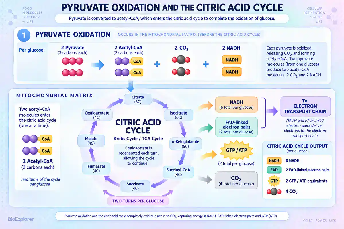 Pyruvate oxidation and citric acid cycle infographic showing two pyruvate converted to acetyl-CoA, followed by two Krebs cycle turns producing NADH, FAD-linked electron pairs, ATP equivalents, and carbon dioxide.
The cycle makes almost no ATP by itself. It does not use oxygen directly. Its job is to strip high-energy electrons from carbon compounds and load them onto carriers.
Stage 4: The electron transport chain and oxidative phosphorylation
Nearly all the ATP appears here. Four complexes sit in the inner mitochondrial membrane. They pass electrons from carrier to carrier and use the released energy to pump protons out of the matrix.
Complex I, also called NADH:ubiquinone oxidoreductase, accepts electrons from NADH and pumps four protons. Complex II is succinate dehydrogenase. It accepts electrons from succinate, and it translocates no protons at all. Complex III releases four protons into the intermembrane space over a full Q cycle. Complex IV oxidizes cytochrome c and passes the electrons to oxygen, pumping two more protons.
Ubiquinone ferries electrons from complexes I and II to complex III. Cytochrome c carries them one at a time from complex III to complex IV. Ten protons per NADH divided by about four protons per exported ATP gives roughly 2.5 ATP per NADH.[15]
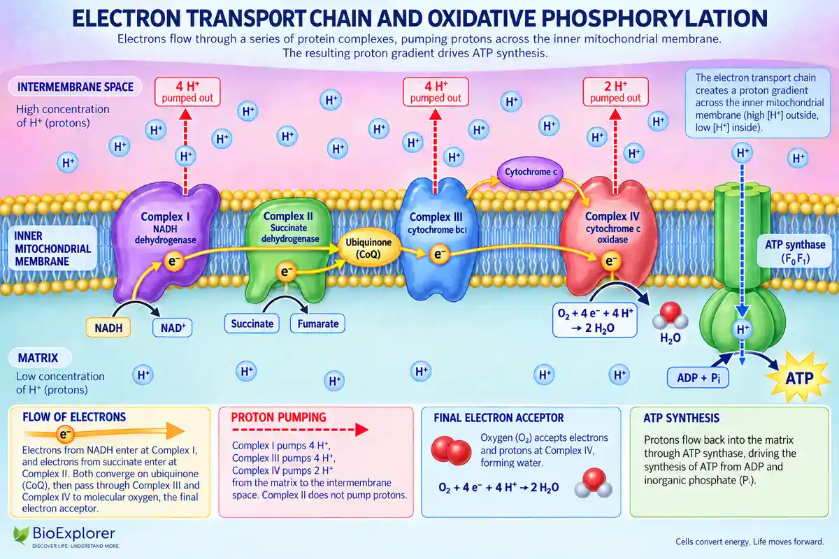 Electron transport chain and oxidative phosphorylation infographic showing mitochondrial Complexes I–IV, CoQ, cytochrome c, proton pumping, oxygen reduction to water, and ATP synthase producing ATP.
A note on what “two FADH2” really means
Tallies commonly say the cycle makes two FADH2 that then enter at complex II. That shorthand is convenient but it misdescribes the chemistry. No free FADH2 molecule travels to complex II. Complex II is the only membrane-bound enzyme of the citric acid cycle. Its SDHA subunit oxidizes succinate and reduces a covalently bound prosthetic FAD group to FADH2, inside the enzyme. Electrons then move through complex II into the coenzyme Q junction.[16]
So “two FADH2” is acceptable bookkeeping for two FAD-linked electron pairs. It is not a description of a diffusing carrier. FAD-linked electrons from fatty acid oxidation are different again. They reach the Q junction through electron-transfer flavoprotein rather than through complex II.
The yield difference now makes sense. Electrons entering at the Q junction skip complex I, which is one of the three proton-pumping stations. Fewer protons cross the membrane, so the pair is worth about 1.5 ATP instead of 2.5.
Chemiosmosis: how a proton gradient becomes ATP
The proton motive force
Pumping protons out of the matrix creates a gradient with two components, one electrical and one chemical. In typical respiring mitochondria the proton-motive force is about 200 millivolts, comprising a membrane potential near 140 millivolts and a pH gradient of about one unit.[17]
The rotor size is not fixed, and neither is the proton cost
ATP synthase has a membrane-embedded rotor ring built from c-subunits. Ring size varies by species. Known F-type rings run from roughly 8 to 17 c-subunits.[18] Mammalian mitochondrial ATP synthase has an eight-subunit ring. With three catalytic sites, that works out near 2.7 protons through the synthase per ATP.
This is why the four protons per exported ATP used earlier is a useful approximation rather than a constant. Ring size differs, so the proton cost of ATP differs, and so does the ATP yield per glucose across organisms.
ATP synthase is a rotary motor
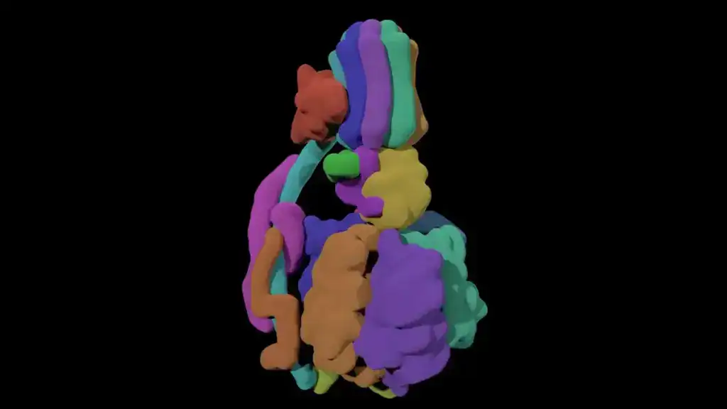
ATP synthase is an enzyme, a molecular motor, an ion pump, and another molecular motor combined. The F0 motor uses the proton gradient to drive the F1 motor.
Protons flow through F0 and spin a circular rotor connected to the F1 head. ADP and phosphate are joined there as it turns.[19] Mechanical rotation is the immediate mechanism of ATP synthesis. The energy source is the proton-motive force that drives the rotation.
The people who worked it out
Peter Mitchell proposed that a proton gradient, rather than a chemical intermediate, couples oxidation to ATP synthesis. The idea was resisted for years. The 1978 Nobel Prize in Chemistry went to Mitchell for formulating the chemiosmotic theory.[20]
The machine itself was described later. Half of the 1997 Nobel Prize in Chemistry went jointly to Paul D. Boyer and John E. Walker, for elucidating the enzymatic mechanism underlying ATP synthesis.[21]
The cycle supplying the electrons was mapped earlier still. The 1953 Nobel Prize in Physiology or Medicine was divided between Hans Adolf Krebs, for the citric acid cycle, and Fritz Albert Lipmann, for coenzyme A.[22]
Where each stage takes place
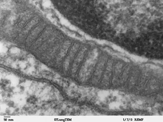
Compartmentation is not a decorative detail. The mechanism depends on a sealed membrane with a proton gradient across it. Location is part of how the pathway works. The table describes typical cases only.
| Stage | Typical mammalian cell | Many bacteria and archaea |
|---|---|---|
| Glycolysis | Cytosol | Cytoplasm |
| Pyruvate oxidation | Mitochondrial matrix | Cytoplasm |
| Citric acid cycle | Mitochondrial matrix, with succinate dehydrogenase in the inner membrane | Cytoplasm, with the succinate dehydrogenase step membrane-associated |
| Electron transport and oxidative phosphorylation | Inner mitochondrial membrane | Cytoplasmic membrane |
Notes on the comparison
Bacteria and archaea lack mitochondria, and they respire anyway. In prokaryotes the enzymes of electron transport and oxidative phosphorylation sit on the inner, or cytoplasmic, membrane.[23]
That membrane does the job the inner mitochondrial membrane does in a eukaryote. Some groups fold it into internal stacks that add surface area. The article on whether prokaryotes have mitochondria covers the arrangement and its evolutionary background.
The tidy version has exceptions inside eukaryotes too. Plant cells run glycolysis in plastids as well as in the cytosol. Some single-celled eukaryotes compartmentalize much of glycolysis in specialized organelles.
A few lineages have reduced their mitochondria to organelles that no longer make ATP. Microbial pathways vary a great deal more than any two-column table can show.
Why the total is 30 in some cells and 32 in others
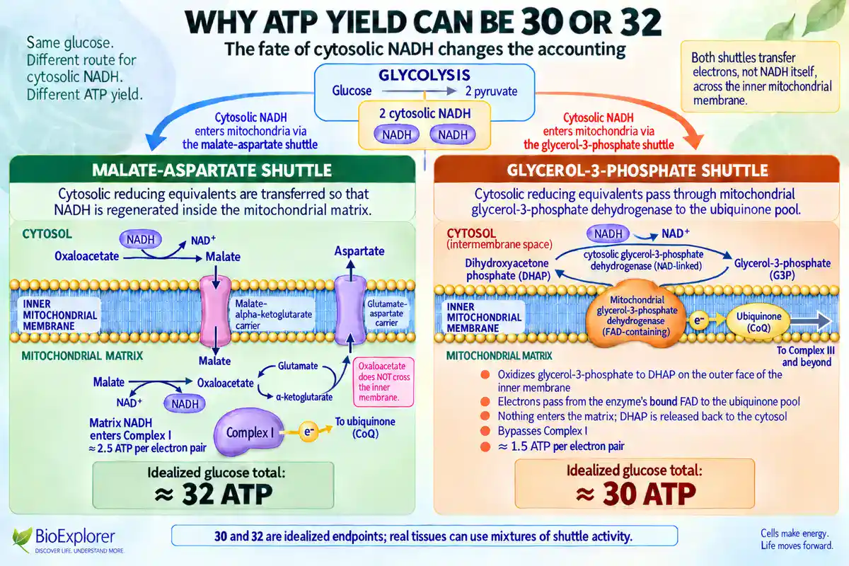
The two NADH from glycolysis are in the cytosol. The inner mitochondrial membrane is not permeable to NADH. A shuttle has to carry the electrons in. Which shuttle operates decides what those electrons are worth.
NADH shuttles move the reducing equivalents of cytosolic NADH into mitochondria. The malate-aspartate shuttle is one of the two major mammalian shuttles.[24] It regenerates NADH inside the matrix. Those electrons enter at complex I and are worth about 2.5 ATP each, putting the total near 32. Tissues differ in which shuttle predominates.
The alternative is the glycerol-3-phosphate shuttle. Its components are expressed in tissues including hippocampal neurons, where it works as a backup route.[25] It hands electrons to the ubiquinone pool through an FAD-containing dehydrogenase, bypassing complex I. Those electrons are worth about 1.5 ATP each, so the total lands near 30.
The 30 versus 32 split is not an inconsistency in the science. It reflects a real difference in how tissues move those electrons. Shuttle use is not strictly either-or, though, so 30 and 32 read best as idealized endpoints rather than fixed totals for any one tissue.
What oxygen actually does
Oxygen is the terminal electron acceptor
Oxygen is not consumed at the start of respiration. It does not react with glucose directly. It waits at the end of the electron transport chain. Complex IV passes electrons to it, and it is reduced to water.
Four electrons and four protons combine with one O2 molecule to form two water molecules. That is why six O2 match the twelve electron pairs carried by ten NADH and two FAD-linked pairs.
Oxygen’s role is thermodynamic. It is the stoichiometric terminal oxidant. Its value to an aerobic cell is its strong affinity for electrons. That makes the drop from NADH to oxygen a long one, and a long drop releases more energy.
What happens when oxygen runs out
Without a terminal acceptor, electrons back up. Carriers stay reduced, proton pumping stops, and the gradient collapses, leaving ATP synthase nothing to run on. NADH accumulates while NAD+ becomes scarce, so the citric acid cycle and pyruvate oxidation stall for want of oxidized carrier.
In a mammalian cell, or in any cell lacking an alternative terminal acceptor, only glycolysis can continue. It continues only if some other reaction regenerates NAD+. Many microorganisms are not in that position, as the section on anaerobic respiration explains.
A poison that proves the point
Cyanide demonstrates the dependency directly. It binds copper and iron centers in the electron transport chain and inhibits cytochrome c oxidase. Oxidative phosphorylation halts, so oxidative ATP production stops. Substrate-level phosphorylation in glycolysis can carry on for a time. Cells shift toward anaerobic metabolism, and lactic acidosis follows.[26]
Oxygen may still be reaching the tissue in cyanide poisoning, but complex IV cannot use it. The block sits on the enzyme that hands electrons to oxygen, not on delivery. That alone is enough to be life-threatening.
Fermentation: keeping glycolysis running without oxygen
What fermentation is for
In homolactic and alcoholic fermentation, the end-product-forming reactions add no ATP. What they do is regenerate NAD+, so that glycolysis can keep operating. Fermentation more broadly conserves energy mainly through substrate-level phosphorylation, and some pathways generate additional ATP after glycolysis. In mammalian cells, when oxygen is limited, pyruvate stays in the cytoplasm and converts to lactate.
Other organisms make ethanol, or one of a wide range of other products, instead. This route is often called anaerobic glycolysis. It nets 2 ATP per glucose, against roughly 32 from complete aerobic oxidation of the same glucose. The reaction regenerates NAD+, which must stay available for the earlier glycolytic reactions to remain favorable.[27]
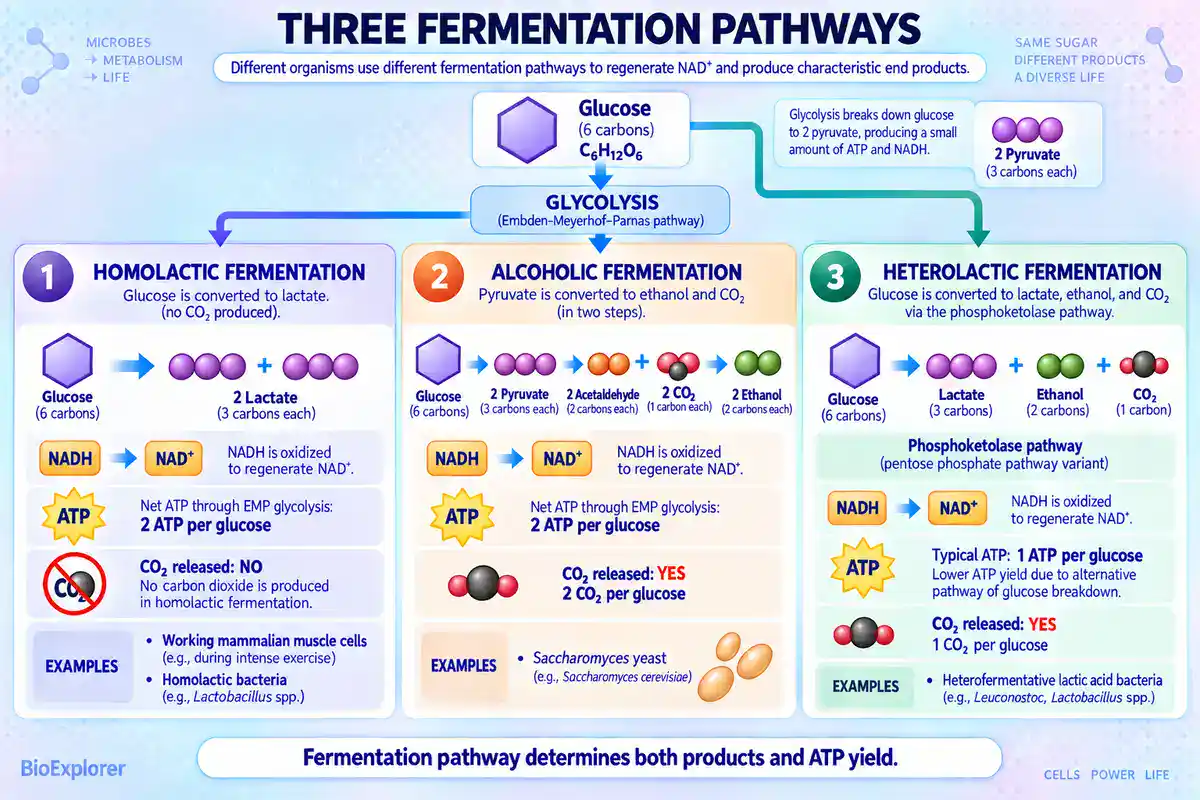
Lactic acid fermentation produces no carbon dioxide
This is the most commonly mangled equation in the topic. Homolactic bacteria metabolize glucose through the standard glycolytic pathway. They yield two lactate molecules per glucose. Lactate dehydrogenase reduces pyruvate to lactate and reoxidizes NADH to NAD+. The net gain is two ATP, and strict homolactic fermentation of glucose generates no CO2.
Heterofermentative bacteria run the phosphoketolase pathway instead. That route does release carbon dioxide. Alcoholic fermentation by Saccharomyces cerevisiae converts sugars into ethanol and CO2.[28]
Written out, the three cases are:
- Homolactic fermentation
- C6H12O6 → 2 lactic acid, plus 2 ATP, no CO2. The same chemistry occurs in working human muscle, where lactate dehydrogenase reduces pyruvate to lactate.
- Alcoholic fermentation
- C6H12O6 → 2 C2H5OH + 2 CO2, plus 2 ATP. This is the yeast route that leavens bread and ferments beer and wine.
- Heterolactic fermentation
- C6H12O6 → lactic acid + ethanol + CO2, plus 1 ATP. This is a bacterial route through the phosphoketolase pathway, not a variant of the muscle reaction.
Two ATP is not a universal fermentation figure
The familiar two ATP belongs to fermentation running through the Embden-Meyerhof-Parnas pathway. Other routes differ, and the differences are not small.
The Embden-Meyerhof-Parnas pathway generates 2 ATP per glucose while reducing 2 NAD+. The Entner-Doudoroff pathway generates only 1 ATP per glucose. The heterolactic pathway also generates 1 ATP per glucose and reduces 3 NAD(P)+.
Meanwhile, conversions of acetyl-phosphate and butyryl-phosphate into acetate and butyrate are coupled to substrate-level phosphorylation, so they generate additional ATP after glycolysis.[29]
Substrate-level phosphorylation is also not the only route. Anaerobic bacteria can use electrochemical ion gradients for ATP synthesis as well, catalyzed by the same H+ or Na+ dependent F1F0 ATP synthases used elsewhere.[30] Those gradients arise without a respiratory chain, through mechanisms such as sodium pumps driven by decarboxylation.
The practical rule is to name the pathway whenever a number is quoted. “Fermentation yields two ATP” is true of homolactic and alcoholic fermentation through the Embden-Meyerhof-Parnas pathway. It is wrong for several other routes.
The page on fermentation in biology covers the applied side of these pathways in food and industry.
Lactate is not a waste product, and it does not cause next-day soreness
Lactate is oxidized as fuel by other tissues. It serves as a gluconeogenic precursor and takes part in cell-to-cell signaling. Dedicated monocarboxylate transporters carry it across membranes rather than letting it simply accumulate. The long-standing assumption that lactic acidosis causes fatigue has been reconsidered against experimental findings that do not support it.[31]
A related belief concerns delayed onset muscle soreness, the ache that arrives a day or two after unfamiliar exercise. Work in the 1980s largely refuted the lactate explanation. Lactate does not linger in muscle on that timescale. The full pathophysiology of that soreness remains unsettled and is still argued over.
Some cells ferment even when oxygen is available
Fermentation is not strictly an emergency measure. Rapidly dividing cells, and tumor cells in particular, convert large amounts of glucose to lactate in the presence of oxygen. Warburg first identified this switch, and it carries his name.
Glycolysis nets 2 ATP per glucose whether conditions are aerobic or anaerobic.[32] Speed and biosynthetic intermediates, rather than ATP per glucose, appear to be what such cells optimize for.
Anaerobic respiration is a different process from fermentation
Membrane-linked electron transfer, without oxygen
Anaerobic respiration uses membrane-linked electron transfer. It generates an ion gradient and makes ATP by chemiosmosis, as aerobic respiration does. During anaerobic growth, electrons can pass to terminal acceptors such as fumarate, nitrate or sulfate, through membrane-bound reductase enzymes.
The resulting ion gradients power cellular processes, including substrate transport and ATP synthesis by membrane-bound ATP synthases.[33]
The terminal acceptor is the most visible difference, but it is not the only one. Respiratory chains also differ in their dehydrogenases, their quinones, their reductases, the ion they couple to, and their proton-pumping stoichiometry. Two anaerobic chains can differ from each other as much as either differs from the aerobic one.
Fermentation is a different arrangement. It uses no respiratory electron transport chain and no external respiratory terminal acceptor. An organic molecule made by the cell takes the electrons instead.
The distinction concerns the respiratory chain rather than membranes in general, because some fermentative organisms do generate an ion-motive force by non-respiratory means.
The confusion between the two is common enough to have been studied. Many undergraduate biology students believe respiration is synonymous with oxygen dependence and organic substrates. The idea that respiration can occur without oxygen, and can involve inorganic substrates, can at first seem alien.[34]
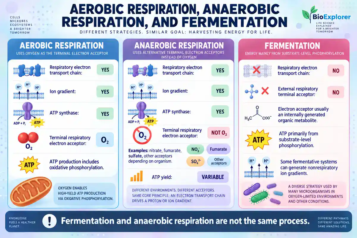
How much energy anaerobic respiration conserves
Anaerobic respiration usually conserves more energy than simple lactate or ethanol fermentation. That is because it retains the ion gradient and the ATP synthase.
The yield cannot be ranked universally against every fermentation pathway. It depends on the electron donor, the acceptor, the organism, and the architecture of the chain. The further electrons fall between donor and acceptor, the more protons can be pumped.
Oxygen is the strongest of the commonly used acceptors, which is why aerobic respiration conserves the most. Beyond that ordering, a single number for anaerobic respiration in general would be misleading.
The categories are less clean than textbooks suggest
Facultative organisms switch between acceptors according to what is available. Escherichia coli has a versatile respiratory chain. It oxidizes ten different electron donors and reduces six different electron acceptors. Salmonella can use two more than that.[35] These organisms use oxygen when present and switch to nitrate or fumarate when it is absent.
The same organism may also do both respiration and fermentation. Pseudomonas aeruginosa is still sometimes described as an obligate aerobe. It has long been known as a facultative aerobe capable of denitrification, using nitrate rather than oxygen as the terminal acceptor. It also grows anaerobically by arginine fermentation and survives anaerobically by pyruvate fermentation.[36]
Obligate anaerobes are a separate category again. Some, such as the methanogens, are genuinely damaged by even small concentrations of oxygen. Using an acceptor other than oxygen and being harmed by oxygen are two different properties. They coincide in some organisms and not in others.
Fuels other than glucose
Fats enter as acetyl-CoA
Fatty acids are activated in the cytosol. Long-chain fatty acyl groups then enter the mitochondrial matrix through the carnitine shuttle, which uses carnitine palmitoyltransferase I and II. Once inside, they are broken down two carbons at a time.
Each round of beta-oxidation yields about 4 ATP equivalents, as one NADH and one FAD-linked electron pair.[37] That pair reaches ubiquinone through electron-transfer flavoprotein and ETF dehydrogenase, not through complex II. Each cycle normally releases one acetyl-CoA, and the final cleavage releases two.
Each acetyl-CoA then enters the citric acid cycle exactly as one derived from glucose would. A long-chain fatty acid goes around many times, so fat yields substantially more ATP per gram than carbohydrate.
Amino acids enter at several points
Amino acids are deaminated first. Their carbon skeletons are converted into pyruvate, acetyl-CoA, or one of the cycle intermediates. Which product forms depends on the amino acid. The nitrogen is disposed of separately, largely as urea in mammals. Protein is a minor fuel under normal conditions. It becomes significant in starvation or prolonged exercise.
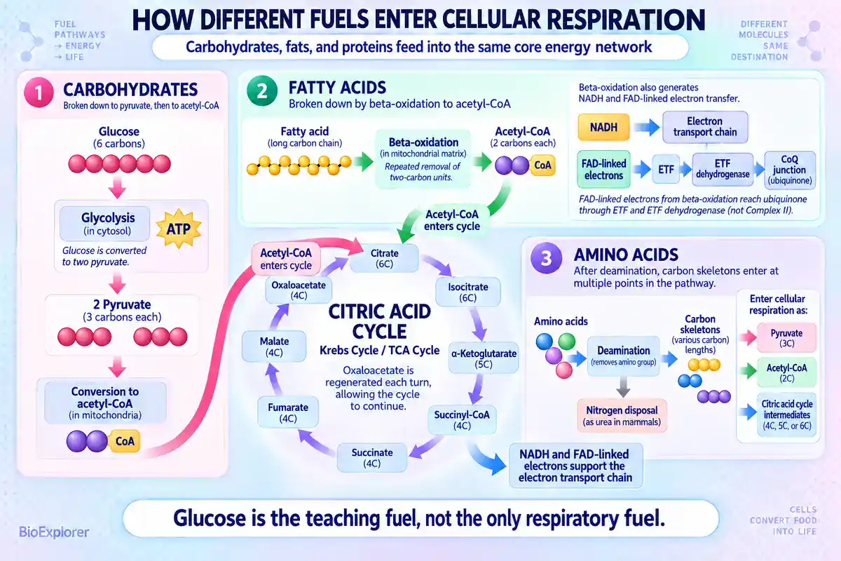
The respiratory quotient reveals which fuel is burning
The fuel in use can be inferred from gas exchange. The respiratory quotient is the volume of carbon dioxide released divided by the volume of oxygen absorbed. It is approximately 1.0 for carbohydrate, 0.7 for fat, and around 0.8 for protein and for a mixed diet.[38]
The value of 1.0 for carbohydrate follows straight from the respiration equation, where six CO2 are produced for six O2 consumed. Fat molecules carry proportionally less oxygen. Oxidizing them consumes more O2 per CO2 released, so the ratio falls.
Cellular respiration and photosynthesis compared
The two processes are often set against each other as opposites. That framing is useful, and loose enough to cause a persistent misconception.
In photosynthesis, energy from sunlight is harvested and used to build glucose from CO2 and H2O. In eukaryotic cells both sets of reactions occur within chloroplasts. The light reactions run in the thylakoid membrane. The carbon-fixing reactions run in the stroma.[39]
Those carbon-fixing reactions are best called the light-independent reactions, or the Calvin-Benson cycle. The older name “dark reactions” is misleading. Several cycle enzymes are themselves light regulated, including fructose-1,6-bisphosphatase and sedoheptulose-1,7-bisphosphatase, whose light-dependent activation is well documented.[40] The reactions do not require darkness and they do not run well in it.
The photosynthesis equation is conventionally written as:
6 CO2 + 6 H2O + light energy → C6H12O6 + 6 O2
| Feature | Cellular respiration | Photosynthesis |
|---|---|---|
| Inputs | Glucose and oxygen | Carbon dioxide, water, light |
| Outputs | Carbon dioxide, water, ATP | Glucose and oxygen |
| Energy | Releases stored chemical energy | Stores light energy as chemical energy |
| Organelle in eukaryotes | Mitochondrion | Chloroplast |
| Carried out by | Plants, animals, fungi, and most microorganisms | Plants, algae, and photosynthetic bacteria |
| Timing | Continuous | Requires light |
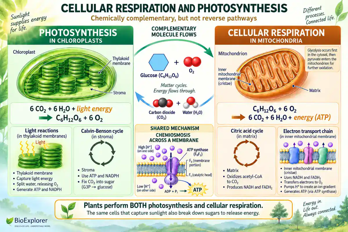
Notes on the comparison
The processes are chemically complementary. They are not simple reversals of each other. They use different enzymes, different organelles, and different routes. Both rely on chemiosmosis across a membrane, which is a genuine shared mechanism.
The important correction is that plants respire. A plant cell runs cellular respiration continuously, day and night, in its mitochondria. It uses sugars its chloroplasts made. In daylight a healthy leaf photosynthesizes faster than it respires. The net exchange then runs one way, which is what makes the misconception easy to acquire.
Why cellular respiration matters outside the classroom
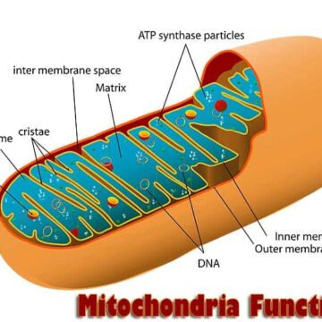
The machinery has a partly separate genetic supply. Mitochondrial DNA is the circular chromosome inside mitochondria, and mitochondria are the organelles that produce ATP.[41]
That description fits human and typical animal mitochondria. Gene content, organization and physical structure vary considerably across other eukaryotic groups.
Several respiratory complex subunits are encoded there rather than in the nucleus. Mutations in that small genome can therefore cause disorders concentrated in tissues with the highest energy demand, such as muscle, heart and brain.
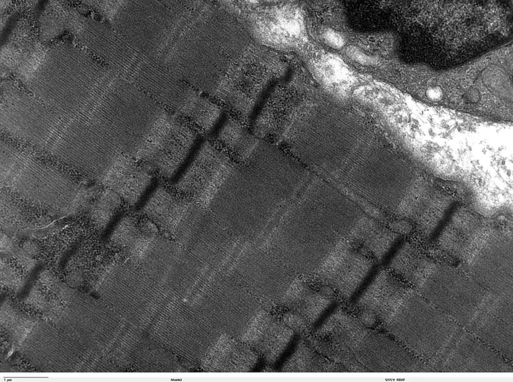
Inheritance of that genome follows its own pattern.
A mitochondrial inheritance calculator models how the proportion of a variant can change between generations. The article on mitochondria functions covers the organelle in more detail.
Common misconceptions
- That aerobic respiration yields 38 ATP per glucose. The figure came from assuming 3 ATP per NADH and 2 per FADH2. Measured proton stoichiometries give 2.5 and 1.5. The modern convention is about 30 to 32 for a mammalian cell.
- That 30 to 32 is a universal constant. It is an idealized figure for a typical mammalian cell. Rotor ring size, shuttle choice, proton leak and coupling state all move it, and plants and microorganisms differ again.
- That fermentation and anaerobic respiration are the same thing. Anaerobic respiration uses membrane-linked electron transfer to an acceptor other than oxygen. Fermentation uses no respiratory chain and no external respiratory acceptor. Many familiar fermentations make ATP mainly by substrate-level phosphorylation, and some fermenters also conserve energy through nonrespiratory ion gradients.
- That every fermentation yields two ATP. Two is right for homolactic and alcoholic fermentation through the Embden-Meyerhof-Parnas pathway. The Entner-Doudoroff and heterolactic routes yield one. Acetate- and butyrate-forming reactions can add more.
- That anaerobic respiration produces only 2 ATP per glucose. Two ATP is a fermentation figure. Anaerobic respiration keeps chemiosmosis and usually conserves more, though the amount depends on donor, acceptor, organism and chain.
- That organisms performing anaerobic respiration die in oxygen. Facultative organisms such as E. coli switch acceptors as conditions change. Oxygen intolerance is a separate trait.
- That lactic acid fermentation releases carbon dioxide. Strict homolactic fermentation of glucose produces lactate and no CO2. Equations pairing lactate with CO2 belong to the heterofermentative pathway.
- That lactic acid causes muscle soreness a day or two after exercise. Lactate does not remain in muscle that long. The lactate explanation was largely refuted decades ago, and the real cause is still debated.
- That free FADH2 travels to complex II. Succinate reduces a covalently bound FAD inside complex II itself. Nothing diffuses there. FAD-linked electrons from fatty acid oxidation arrive by a different route again.
- That plants photosynthesize and animals respire. Plants do both. Their mitochondria run continuously, and in darkness a plant is a net consumer of oxygen.
- That cellular respiration is the same as breathing. Breathing moves air in and out of lungs. Cellular respiration is chemistry inside every cell, and organisms with no lungs perform it.
- That mitochondria are required for cellular respiration. Bacteria and archaea have none and respire on their cytoplasmic membrane.
- That the citric acid cycle uses oxygen. No step of the cycle reacts with O2. In a typical mammalian cell it stops without oxygen, because the chain stops regenerating NAD+ and FAD. Organisms that respire anaerobically regenerate those carriers without oxygen and keep the appropriate cycle reactions running.
- That glycolysis requires oxygen. Glycolysis is cytosolic and needs no oxygen. The reaction sequence is the same either way, although its flux, its regulation and the fate of its pyruvate are not.
- That ATP stores energy in its phosphate bonds. Breaking a bond costs energy. ATP hydrolysis is favorable because the products are more stable overall, which is what gives ATP its high phosphoryl-transfer potential.
Frequently asked questions
C6H12O6 + 6 O2 → 6 CO2 + 6 H2O + energy. One glucose molecule and six oxygen molecules produce six carbon dioxide molecules, six water molecules, and energy captured as ATP. The equation summarizes dozens of enzyme-catalyzed reactions.
About 30 to 32 ATP per glucose in a typical mammalian cell. Four come directly from substrate-level phosphorylation in glycolysis and the citric acid cycle. The other 26 to 28 come from oxidative phosphorylation, using about 2.5 ATP per NADH and 1.5 per FAD-linked pair. The figure is an idealized convention rather than a fixed constant.
Older sources assumed each NADH yields 3 ATP and each FADH2 yields 2. That gives 38, or 36 with the less efficient shuttle. Those ratios were estimates that measurement did not support. Proton-based values of 2.5 and 1.5 are now standard, and they lower the total.
Glycolysis in the cytosol splits glucose into two pyruvate, netting 2 ATP and 2 NADH. Pyruvate oxidation in the matrix converts each pyruvate to acetyl-CoA, releasing CO2 and NADH. The citric acid cycle oxidizes the acetyl group completely. The electron transport chain and oxidative phosphorylation then make most of the ATP.
In a typical mammalian cell, glycolysis occurs in the cytosol and the other three stages in the mitochondrion. The electron transport chain sits in the inner mitochondrial membrane. In many bacteria and archaea, which have no mitochondria, glycolysis and the cycle run in the cytoplasm and electron transport runs on the cytoplasmic membrane.
Oxygen is the terminal electron acceptor. It sits at the end of the chain, takes electrons from complex IV along with protons, and is reduced to water. It does not react with glucose directly. Its role is thermodynamic: a strong affinity for electrons makes the drop from NADH a long and productive one.
Both use membrane-linked electron transfer and make ATP by chemiosmosis. Aerobic respiration ends with oxygen. Anaerobic respiration ends with something else, such as nitrate, sulfate, fumarate or carbon dioxide. The chains also differ in their carriers and reductases, not only in the final acceptor.
No. Fermentation uses no respiratory electron transport chain and no external respiratory terminal acceptor. Its role is to reoxidize NADH so that glycolysis can keep running. Many familiar fermentations make ATP mainly by substrate-level phosphorylation, mostly within glycolysis, although some pathways add more at the product-forming steps. Some fermenters also conserve energy through nonrespiratory ion gradients that drive ATP synthase.
Two ATP per glucose for homolactic and alcoholic fermentation running through the Embden-Meyerhof-Parnas pathway. The Entner-Doudoroff and heterolactic routes yield one. Some acetate- and butyrate-forming pathways generate additional ATP after glycolysis. The pathway has to be named for the number to mean anything.
C6H12O6 → 2 CH3-CHOH-COOH, with a net gain of 2 ATP. No carbon dioxide is released. Equations showing lactate together with CO2 describe the heterofermentative bacterial pathway, which runs through different enzymes.
They are chemically complementary. Photosynthesis stores light energy by building glucose from carbon dioxide and water, releasing oxygen. Cellular respiration releases that energy by oxidizing glucose back to carbon dioxide and water. They are not reverse reactions, and plants carry out both.
Yes, continuously, in mitochondria, using sugars made during photosynthesis. In daylight photosynthesis usually outpaces respiration in a healthy leaf, so net gas exchange runs toward oxygen release. In darkness only respiration continues, and the plant is a net consumer of oxygen.
Close to half of the energy theoretically available from oxidizing glucose is captured in ATP. The exact percentage depends on the free-energy convention used and on how tightly the mitochondria are coupled, so it should be read as an approximation. The rest is released as heat, which helps endothermic animals stay warm.
Life is so complex.
Cite this page
BioExplorer. (2026, September 8). Cellular Respiration Equation: Stages, Products, and ATP Yield. https://www.bioexplorer.net/cellular-respiration-equation.html/
About the author

MSc Genetics & Cytology
Anastasiia Nesterenko holds an MSc in Genetics and Cytology and spent sixteen years as a research assistant at the Mechnikov Institute for Microbiology and Immunology. She has written for BioExplorer since 2016, from genetics and immunology to primates, insects, plants and flowers.

this was a really good article thanks for making this