Cell Biology Discoveries in 2019: In 2019, researchers managed to decipher multiple complex mechanisms that underlie the functioning of the cells of the body.
While the Nobel Prize of 2019 was awarded to the group of researchers that have found components of the oxygen-sensing system in the cells, current research has found novel mechanisms that lie based on other crucial aspects of cell life.
For example, multiple researchers have focused on the mechanisms of cell division, such as attachment of the mitotic spindle to the chromosomes and the formation of the microtubules.
Another crucial area that attracts multiple researchers is the biology of stem cells and cellular reprogramming. One of the crowning achievements of this branch of cellular biology is the development of functional blastocysts from stem cells in mice, which has multiple implications for science.
History of Cell Biology
Other advancements in cell biology may not be as staggering, but can still contribute significantly to the development of fundamental science and medicine. Let us discover some of them.
Table of Contents
- Top Cell Biology Discoveries in 2019
- Surprising secrets of hair follicles: a certain type of hair follicle stem cells can differentiate into neurons and glial cells [USA, April 2019]
- Reprogramed cells for diabetes relief: a new approach in mice can point to the potential diabetes treatment [Switzerland, February 2019]
- Immune cells and diabetes: a new type of immune cell discovered in type I diabetes patients [USA, May 2019]
- There is more to cell division: new insights into cell division mechanisms have come to light [UK, March 2019]
- A small, but the important secret of the telomeres: researchers found a mechanism for telomere maintenance [France, 2019]
- Building backward: a team of international investigators has deciphered the makeup of microtubules with the help of reverse engineering approach [USA, May 2019]
- How cells form protective bags: the researchers have uncovered proteins responsible for changes in the plasma membrane under stressful conditions [Spain, December 2019]
- Back to the early embryo: the researchers have created artificial structures typical for early embryonic development from reprogrammed mouse stem cells [Japan, August 2019]
- It is not easy to reach the oocyte: the researchers have found a protein crucial for normal movement of the sperm [Japan, December 2019]
- Nobel Prize 2019 Winners: William G
- Top 15 Discoveries in Cell Biology for 2018
Top Cell Biology Discoveries in 2019
Here are the top 10 recent discoveries in cell biology 2019.
Surprising secrets of hair follicles: a certain type of hair follicle stem cells can differentiate into neurons and glial cells [USA, April 2019]
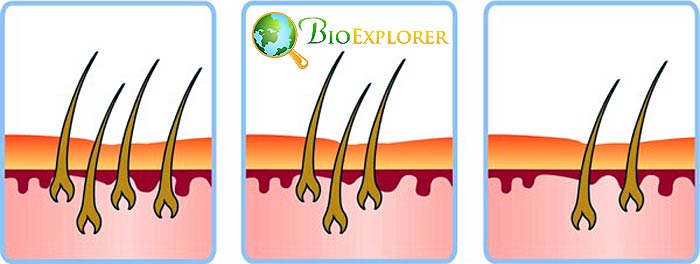
The development of the hair follicle is a complex process with several stages. While studying hair follicle development in mice, the team led by Hornyak T. J.has found a cellular subset with unexpected properties.
- The hair follicle development has three distinct stages: anagen, catagen, and telogen.
- Telogen is the stage when melanocyte stem cells do not divide and enter a “rest period“.
- The researchers have found two parts of the follicles with distinctly different melanocyte stem cells populations.
- One area was called the secondary hair germ compartment and contained cells that were fated to become hair melanocytes. These cells lacked CD34 marker.
- Another compartment contained CD34+ cells. These cells had a genetic profile similar to stem cells found in the neural crest.
- Further research has shown that these particular cells could produce myelin that protects neurons in certain conditions.
This finding shows that hair follicles contain very diverse cell types. It may be necessary for helping scientists understand how some types of cancer develop.
![]()
Reprogramed cells for diabetes relief: a new approach in mice can point to the potential diabetes treatment [Switzerland, February 2019]
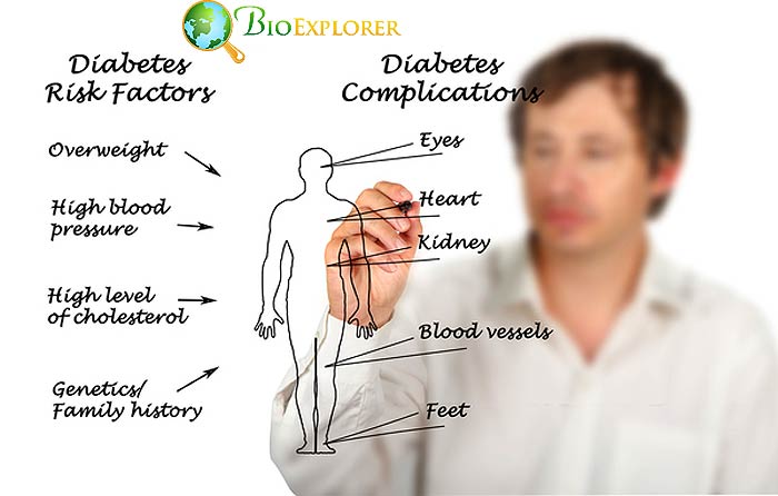
Certain animals can change the identity of the differentiated cells in response to stress. It is known that in mice, certain types of cells that used to produce glucagon or somatostatin in the pancreas could be reprogrammed into insulin-producing cells when regular β-cells of the islets are destroyed.
A team under the leadership of Professor Pedro Herrera at the University of Geneva has tried to re-create this reprogramming process in human cells.
- The researchers took pancreatic islet cells that did not produce insulin from deceased human donors (α- cellsand cells that produce pancreatic polypeptide).
- The cells could be reprogrammed using transcription factors PDX1 and MAFA.
- The reprogrammed cells produced insulin in response to the presence of glucose.
- If the reprogrammed cells are transplanted into diabetic mice, their presence reverses diabetes, and the positive effect was preserved for six months.
- The reprogrammed cells still had some features of their previous lineage.
This study provides new insight into the plasticity of cells. It could help with the development of treatments both for diabetes and diseases that involve the destruction of specific cell types.
![]()
Immune cells and diabetes: a new type of immune cell discovered in type I diabetes patients [USA, May 2019]

Our immune system has two major cell types: T-cells and B-cells, each with its own array of properties. The team of researchers at the John Hopkins University School of Medicine has found that patients with diabetes type I have slightly different immune cell composition:
- Type I diabetes patients have an immune cell type that combines features of both B and T-cells.
- A majority of this double – type cells contain a previously unknown receptor.
- This receptor binds with HLA-DQ8 strongly.
- HLA-DQ8 is a molecule that can bind insulin and present it to T-cells.
- If the HLA-DQ8 – insulin complex binds to T cells, the T-cells may be primed to attack insulin-producing cells.
- The receptor discovered in the novel immune cell type can act as an especially potent stimulator of anti-insulin T cells.
The researchers think that these cells may drive the development of type I diabetes. These cells could potentially be used as a marker of early disease development in diabetes type I patients.
Still, there are many questions about these new cells that are not answered yet.
![]()
There is more to cell division: new insights into cell division mechanisms have come to light [UK, March 2019]
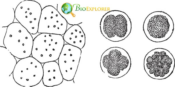
Cellular division is controlled by two families of enzymes: kinases and phosphatases. Kinases are responsible for activating processes, while phosphatases are blocking them.
Phosphatases are significantly less researched than kinases. A team led by A. Saurin at the School of Medicine of the University of Dundee has studied a phosphatase family PP2A-B56.
- PP2A-B56 is a complex protein that attaches phosphate groups to aminoacids serine and threonine.
- The chromosomes have two crucial areas that are important during cellular division:
- Centromere – a structure that unites two chromatids of the chromosome
- Kinetochore – an area where the spindles attach to move chromosomes during division.
- There are several forms of PP2A-B56 that are localized either at the centromere of the chromosome or at the kinetochore.
- As a result, these proteins control 3 crucial processes:
- The attachment of sister chromatids in the centromere area.
- The attachment of microtubules to the kinetochore.
- The assembly of the division spindle.
Recently, inhibition of specific proteins in the PP2A-B56 family was shown to be helpful in the treatment of neurodegenerative diseases.
Understanding how specific forms of PP2A-B56 work in a cellular division may help with the development of better-targeted agents that could stop a particular cause of disease.
The inhibition of PP2A-B56 may also be used in cancer treatment.
![]()
A small, but the important secret of the telomeres: researchers found a mechanism for telomere maintenance [France, 2019]

Telomeres are structures located at the ends of chromosomes. Though the sequences in the telomeres do not code for any known genes, they still play an important role. The presence of telomeres helps prevent the destruction of chromosomal DNA underneath.
That is why the shortening of telomeres is linked to aging, while lengthening of the telomeres often happens in cancer cells. The replication and maintenance of telomeres is a complex process.
One of the proteins responsible for telomere maintenance is called CST. This protein is present in all cells from yeast to mammals.
Mutations in the genes responsible for the CST protein can lead to a dangerous syndrome in humans that is associated with disorders in multiple organ systems, including the brain, bones, and gastrointestinal tract.
An international team of researchers from France and Portugal has studied a part of this complex called Stn1 in fission yeast Schizosaccharomyces pombe.
- Stn1 is controlled by a phosphatase ssu72.
- When ssu72 adds phosphorus to Stn1, the protein prevents the elongation of the telomere.
- If another protein, Cdk1, attaches to Stn1, then the latter cannot attach to the telomere, and new sequences are added to telomerase.
- The same mechanism of telomerase regulation is present in mammalian cells.
It is vital to understand how the telomeres are maintained, as telomerase is an important protein in cancer development, as well as in aging.
![]()
Building backward: a team of international investigators has deciphered the makeup of microtubules with the help of reverse engineering approach [USA, May 2019]
.
When cells divide, a specialized structure is formed called mitotic spindle. This spindle is composed of microtubules – complex structures that are composed of several components. Microtubules originate in several spots:
- From the so-called mitotic spindle poles.
- From the chromosomes
- By branching from the surfaces of already formed microtubules.

The exact steps of branching of microtubules were previously unknown. Three researchers at the Princeton University – Sabine Petry, specializing in molecular biology, Howard A. Stone, specializing in engineering, and Joshua W. Shaevitz, a physics professor – have collaborated to reconstruct this process.
- The team has used eggs of the Xenopus frog as a model.
- They have established that new microtubules tend to form on the surface of the pre-existing ones.
- The researchers have regulated the availability of the three factors that regulate microtubule growth: TPX2, augmin, and γ-TuRC.
- The experiments have shown the following sequence:
- TPX2 is the first to be deposited on the surface of the “old” microtubule.
- Augmin and γ-TuRCare bound together.
- The new microtubule branches are formed.
- Based on the results of the experiments, a computer model of the microtubule branching was developed.
This work was unique as it required the collaboration of specialists from three different fields. It is also crucial for understanding one of the most important questions of cell biology – cellular division.
Sabine Petry, one of the researchers, has received an WICB Junior Award for Excellence in Research for her work.
![]()
How cells form protective bags: the researchers have uncovered proteins responsible for changes in the plasma membrane under stressful conditions [Spain, December 2019]
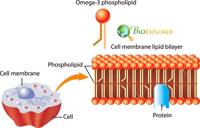
Some cells in our bodies spend their lives in constant danger. For instance, muscle cells are subject to regular stretching, skin cells often fall victim to mechanical pressure, other cells can find themselves in adverse chemical conditions. To withstand such stresses, cells have several methods of defense:
- The cellular membranes form “bags ” called caveolae to withstand pressure.
- In some cases, caveolae can form clusters that are called caveolae rosettes.
- The cells form so-called stress fibers that help the membrane to bend.
A team of researchers has studied the molecular underpinnings of caveolae formation:
- They used the culture of human skin cells for their experiments.
- To create stressful conditions, the cells were either subjected to stretching or were placed in conditions with lower osmotic pressure.
- The experiments have shown that caveolae are formed with the help of a protein called FBP17.
- FBP17 attaches to the caveolae to arrange them in caveolae rosettes.
- Another protein, c-Abl, regulates the activity of FBP17.
- c-Abl senses mechanical pressure and blocks the activity of FBP17.
- Instead of changing the curve of the membrane and formation of caveolae, c-Abl helps release the stress fibers that help withstand mechanical tension.
In people with muscular dystrophies, these stress-relieving mechanisms are impaired. By understanding the mechanisms that help the cells cope with stress, it could be possible to treat people with these severe conditions.
![]()
Back to the early embryo: the researchers have created artificial structures typical for early embryonic development from reprogrammed mouse stem cells [Japan, August 2019]
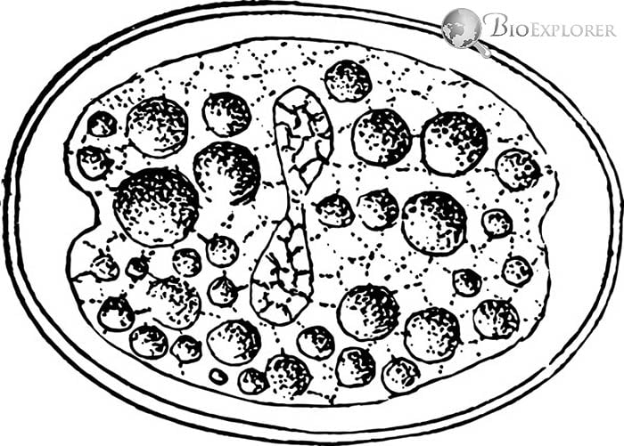
Right after fertilization, the zygote divides into two cells. Those two cells are called totipotent – they can differentiate into any possible type of cell.
Totipotent cells can potentially grow into the whole embryo, while stem cells do not have this ability and have a certain limit.
Japanese scientists have tried to reprogram differentiated cells into totipotent cells – which have never been achieved before:
- In the course of the experiment, the scientists used mouse pluripotent stem cells.
- The cells underwent a complex reprogramming process, including adding transcription factor Prdm14.
- After reprogramming, the cells self-organized into structures that were similar to a blastocyst stage of embryonic development.
- Blastocysts are structures with an outer structured layer, and a cavity with a mass of undifferentiated cells within that later give rise to the embryo.
- Artificial blastocytes had a similar structure, and the mass of the cells inside their cavities was pluripotent.
- The researchers have implanted the blastocysts into mice.
- The implanted artificial blastocysts could temporarily grow inside the uterus.
This achievement could help scientists understand what factors make cells totipotent. These bits of knowledges are essential to several fields of medicine.
![]()
It is not easy to reach the oocyte: the researchers have found a protein crucial for normal movement of the sperm [Japan, December 2019]

The cells have a complex system of proteins. Some of them are responsible for shuttling ions in and out of the cell, thus creating an electrical charge. These ion transporter proteins are in turn controlled by another group called phosphoinositides.
It is known that both types of proteins are present in the sperm cells, and defects in the production of these proteins may lead to male infertility.
A team of Japanese researchers was interested in the role of electricity-related protein in the sperm.
- They have focused on the VSP – voltage sensing protein that was known to be present in several of organs, including the testes.
- The researchers have found that VSP is present in the sperm as well.
- Sperm without VSP was not able to reach the oocyte properly because it was swimming in circles.
- It was established that VSP controlled the distribution of certain phosphoinositides in the sperm that were crucial for normal sperm orientation and motility.
This discovery can help develop new treatments for male infertility.
![]()
Nobel Prize 2019 Winners: William G

Oxygen is crucial for the normal functioning of cells. Therefore, the studies devoted to various roles of oxygen in the cells were awarded the Nobel Prize through the years. In the year 2019, the award went to the three researchers – William G. Kaelin Jr. , Sir Peter J. Ratcliffe, and Gregg L. Semenza. This trio has discovered another piece of this puzzle: how cells can sense the amount of available oxygen. Each of these scientists has contributed to the discovery of oxygen-sensing mechanism:
- It was known at the beginning of the 20th century that when the oxygen levels in the body are low, the kidney cells start producing a hormone called erythropoietin (EPO).
- Erythropoietin stimulates the development of new erythrocytes in the blood that can transport more oxygen to the cells of the body.
- Gregg Semenza has studied the regulation of erythropoietin gene in response to different oxygen levels.
- In course of his experiments, Semenza has discovered a protein called hypoxia-inducible factor.
- This factor binds to regulatory segments of the EPO gene in response to a reduction in oxygen levels.
- Semenza has also discovered the genes responsible for the HIF components, HIF-1α and ARNT.
- William Kaelin Jr. was studying the VHL gene and has discovered that this gene codes for a protein that controls responses to hypoxia.
- When VHL is absent in the cells, many hypoxia -linked genes are activated, and it can lead to the development of certain cancers.
- Sir Peter Ratcliffe has found that VHL interacts with HIF-1α, a part of the HIF complex, and induces its degradation when oxygen levels are normal.
- Later, both Kaelin and Ratcliffe have discovered prolyl hydrolases, specialized enzymes that mark HIF-1α fordestruction in the presence of normal oxygen levels.
All three discoveries were crucial in our understanding of how cells react to various oxygen levels. As this mechanism is disrupted many of conditions from infections to cancer and infarction, this research was crucial for medicine and fundamental research alike.
![]()
The year of 2019 has shown how much we actually do not know about the inner workings of the cell. For example, though not presented in the current article, there were multiple experiments devoted to the formation and activity of microtubules: from the physical forces driving their movement to the fact that microtubules may contribute to diabetes development.
Other laboratories devoted their work to previously overlooked structures, such as cilia in kidney cells or flagella of the archae a. As we can see, new types of cells are found in unexpected situations – such as hair follicles or immune system in diabetes patients.
Top 15 Discoveries in Cell Biology for 2018
All recent advances in cell biology points to one conclusion – it would be impossible to find remedies for many known diseases unless we would understand the smallest units of our bodies – the cells – better.
![]()


