From the scientific point of view, the year 2020 was devoted to public health, vaccine, and virus research. Still, without knowing the details of our anatomy and physiology, it could be impossible to understand the influence of any illness on the body, including COVID19.
Despite all the drawbacks the pandemic has caused, some outstanding research was published in Anatomy and Neuroscience. Brain anatomy, development, and disorders are gaining considerable interest from researchers, which is also reflected in the newsworthy discoveries and developments in this area.
Top Anatomy News in 2020
This list of the most noteworthy Anatomy discoveries in 2020 shows that even in dire times, it is imperative to understand one’s body in the first place to fight the challenges our environment currently throws our way.
1. Fibers inside the heart are crucial for its normal function – Great Britain, August 2020.
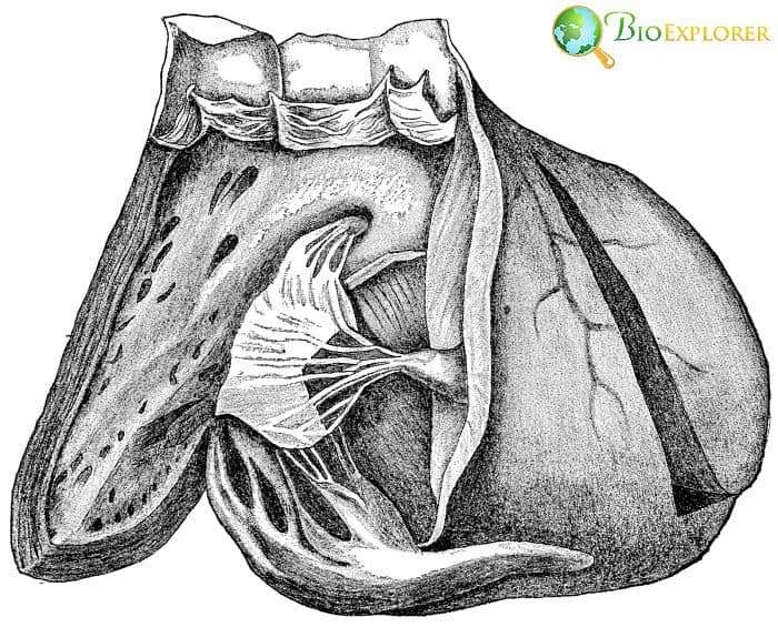
Sometimes, the ancient knowledge obtained many centuries ago can gain new meaning and significance in the present time. This has happed to specific heart structures. The heart as an organ was fascinating for many people even before anatomy has become an acknowledged science.
For instance, Leonardo da Vinci, a famous painter, and inventor has dissected hearts and has found a previously unknown structure called a moderator band or a septomarginal trabecula. This band of fibers is located in the heart’s right ventricle. These trabeculae appear in the heart when it just begins developing in the fetus.
Previously, it was thought these structures were just something that remains present from the early development. The scientists from the Imperial College of London have shown it was not so:
- The team has done genome-wide sequencing in more than 18000 participants to find genes linked to the inner heart structure and the flow of blood in the cardiovascular system.
- The researchers also searched for the links between specific genes, anatomical structures of the heart, and various cardiac diseases.
- The scientists also included the inner fibers of the heart into their heart models. They looked at the way the inner heart “branches” possibly influence heart function.
- Scientists have found that the heart’s trabeculae can influence the heart’s electrical activity and its ability to pump blood properly.
- A specific arrangement of the heart fibers may predispose patients to potential heart failure.
- By analyzing heart structure, it could be possible to find people potentially at risk for heart disease.
Suggested Reading:
Best Anatomy and Physiology Coloring Workbook Review
![]()
2. A new type of salivary gland found – Netherlands, October 2020
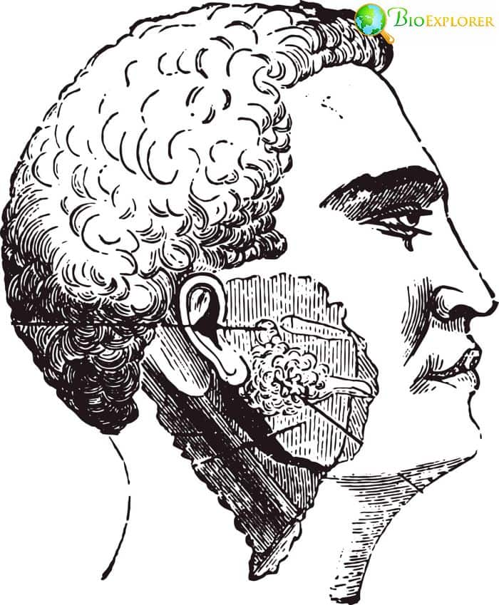
Humans have multiple glands that are located in the tissues of our airways and digestive tract. There are three critical glands, and there are also multiple minor glands hidden in the upper airways and digestive tract.
These glands have multiple functions, from producing saliva, which aids in digestion, to filter potentially harmful agents as part of the immune system “firewall“.
Recently, a group of researchers at the Netherlands Cancer Institute used a new testing approach for making scans in the head and the neck area. They have found something unexpected:
- The research team has combined positron emission tomography/computed tomography using special chemicals that bind to the cells called ligands for the prostate-specific membrane antigen (PSMA).
- These chemicals have radio labels attached and so can help visualize structures during tomography scans.
- When applying the new approach, scientists have found a previously unseen pair of salivary glands called tubarial glands.
- Such glands were found in approximately 100 cancer patients.
- The inner anatomy of this new pair of organs was similar to other salivary glands.
- Tubarial glands were found to be very sensitive to radiotherapy.
- Damage to these glands can lead to problems with swallowing and producing saliva.
- The scientists recommend evading them during radiotherapy procedures.
- There is currently a debate about this discovery.
- Some specialists point out that a similar type of glands was known since the previous century.
- Still, the fact that these glands can influence the outcome of cancer treatment and quality of life in cancer patients is a discovery in itself.
Suggested Reading:
Top 25 Urinary System Fun Facts
![]()
3. The classification of Parkinson’s disease development awarded first Parkinson’s disease prize – Netherlands, July 2020.
Parkinson’s disease is a severe disorder of the nervous system. The leading cause of Parkinson’s is the loss of nerve cells in the brain called substantia nigra. As this area is responsible for movement control, the patient develops tremors and then progressively loses the ability to perform coordinated movements.
As this disease is devastating to patients and is impossible to cure, this syndrome’s mechanisms and possible treatment options focus on research for many scientists.
This year 2020, the first prize for best research related to this topic was awarded by the Journal of Parkinson’s disease. The prize was given to a team of scientists from Germany that have documented how the brain changes during this disease, dividing the changes into stages.
- To document the changes, the researchers used samples taken from various brain areas that were unusually thick and therefore contained multiple cell types.
- Scientists have proven that there are 6 main stages of Parkinson’s disease development.
- The disease progresses as certain nerve cell types accumulate so-called Lewy bodies made of the protein called α-synuclein.
- The group has shown for the first time that these Lewy bodies could first appear very far from substantia nigra – namely, in the nerve system of the nose and the intestine.
- The Lewy bodies appear in the substantia nigra cells in later stages of the disease.
- These findings have provided the specialists with the information that allowed them to find markers of the early stage of the disorder.
- This year, another team of scientists has confirmed what the earlier research suggested – the early stages of Parkinson’s disease can indeed start in the gut.
Suggested Reading:
25 Mind-Blowing Biology Breakthroughs That Shaped Our World!
![]()
4. Early brain development recreated in cell culture – Denmark, May 2020

It is hard to study the human brain at the early stages. There are simply too few samples to use as brain development occurs in the early stages of pregnancy.
A team of researchers from Denmark has decided to look outside the body instead of trying to get inside:
- During their experiments, the researchers have used a culture of human stem cells.
- The cells were placed in an environment with several specific molecular signals that direct cell fate.
- Such an environment mimics the situation in the developing egg.
- The stem cells’ culture has transformed into a nerve tissue with areas corresponding to developing brain areas – hindbrain, midbrain, and forebrain.
- With this new model, it is possible to chart the stages of brain development in great detail.
- It is possible to use genetic typing to identify the cells composing each developing brain region.
- This invention can bridge brain anatomy, genetics, and stem cell therapy.
- The model can help medical professionals develop specific stem cells to treat various disorders connected to nerve cell death.
Suggested Reading:
Top 15 Anatomy & Physiology News In 2019
![]()
5. The brain is less adaptable than previously thought – December 2020, Sweden
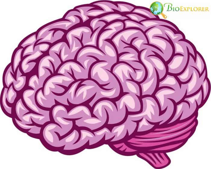
For many years, specialists thought that the brain can reorganize itself in serious injuries, such as losing a limb. With new bionic prostheses, this prevailing theory has come under scrutiny.
- Modern prostheses can be linked to the nerves in the remaining parts of the amputated limb to make the new “arms” similar to the “real ones“.
- The surgery is still far from perfect. Often, the surgeons can not find the exact nerve endings for each type of movement or perception.
- The research team from Sweden has found that this problem translates to misplaced nerve signals.
- For example, when a person touches an object with a thumb, they feel the touch in another area of the hand.
- This misplaced feeling does not shift even with constant exercise with the new bionic arm for prolonged periods.
- This means that the brain is not as adaptable – the areas of the brain responsible for receiving touch information do not get reorganized to process the information received from the replacement arm correctly.
- This finding corrects the predominant myth of brain plasticity. It helps make a more realistic picture of what our nervous system is capable of.
Suggested Reading:
Top 25 Fun Facts About The Skeletal System
![]()
6. Lymph vessels in the liver can cause plastic bronchitis in vulnerable patients – USA, December 2020.
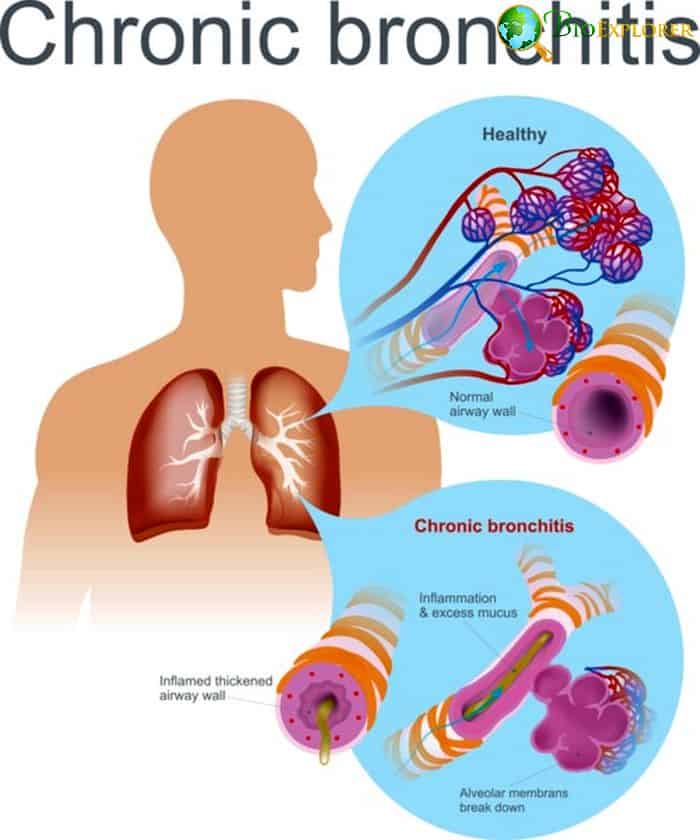
Some complicated heart surgeries have unexpected side effects. For instance, children with Fontan surgery are at risk of developing plastic bronchitis.
This particular illness is caused by the unusual lymph flow that leads to the lymph getting into the lungs and causing so-called casts.
- To treat plastic bronchitis, it is imperative to find how exactly the lymph gets into the lungs in the first place and block its road.
- A group of radiologists at the Nemours/Alfred I. duPont Hospital for Children have had a child patient with a case of plastic bronchitis.
- Traditional methods have not helped in finding the source of lymph in their lungs.
- By applying a new method for imaging the lymph vessels called dynamic contrast-enhanced magnetic resonance lymphangiogram, the radiologists have found the source of the lymph in the complex net of lymph vessels in the liver.
- Thanks to their finding, they could search for a similar vessel in other patients with a similar problem. Another case was already treated thanks to their discovery.
- This is an important finding that can assist medical specialists in treating plastic bronchitis patients in the future.
Suggested Reading:
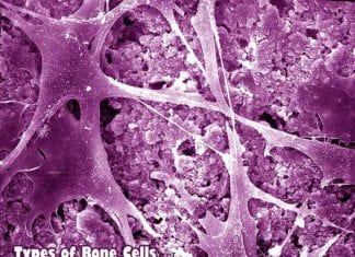
Types of Bone Cells
![]()
7. Nanoparticles show new aspects of bone structure – Denmark-Sweden-Switzerland-France, June 2020.

It is essential to study bone structure to treat illnesses affecting our limbs and spine.
- A collaboration between the scientific institutions and various specialists across several European countries -Denmark, France, Sweden, and Switzerland was created to study the microscopic structure of bones.
- The combined team of physicists, chemists, and biomedical specialists used novel X-ray methods to study nanocrystals in the bones.
- Bones are composed of collagen fibrils and mineral content that usually create a layered, uniform structure.
- The new approach used by the multidisciplinary team has found that there are areas absent in conventional models on the level of these nanoparticles.
- Currently, it is not clear how valuable this information is. Hopefully, it would help revise the existing bone structure models and assist in making better prostheses.
Suggested Reading:
Top 10 MCAT Prep Books – An Ultimate Guide
![]()
8. We can walk upright because our feet are stiff – USA-Japan-UK, February 2020

If one looks at great apes, one could notice that their feet are flat and flexible. Our feet, on the other hand, have less flexibility and are arched in the middle. A team of scientists has shown the importance of this arch in our feet:
- An international team of scientists has looked into specific components of the human feet that contribute to this stiffness.
- The team has discovered that the transverse tarsal arch formed by the bones in the foot interacts with the tissues inside the foot itself, creating a stiff supporting structure.
- This complex structure helps us have such various movements, from a leisure walk to a fast run.
- The bones inside the human foot can be arranged differently, which can influence the overall stiffness and the curve of the arch.
- It may potentially influence movement as well.
- The researchers have also discovered that the curve of the arch has changed through human evolution.
- According to the fossil record, feet with the curved foot arch have appeared even before the appearance of our genus Homo.
- This finding is essential for several crucial applications:
- Treatment of syndromes associated with flat feet.
- Creation of robotic feet and prostheses.
- Study of foot function in general.
Suggested Reading:
Top 25 Endocrine System Fun Facts
![]()
9. Males and females have different brain structures – USA, August 2020.
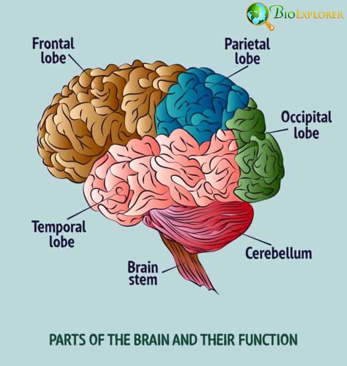
The debate whether there are differences between how the brains of women and men function are always on.
A large study conducted by S. Liu and colleagues at the National Institute of Neurogenomics at Bethesda, Maryland, seems to bring new, compelling arguments to this discussion:
- Studies in mice have shown that the brain’s gray matter is spread differently across the brain regions in male and female mice.
- The researchers have decided to use the data collected for the Human Connectome Project (HCP), which included 976 adults without any known diseases ranging between 22 and 35 years, to test whether similar features are typical for human brains.
- The analysis has revealed that the content of gray matter in a particular area is different between sexes.
- In women, the areas responsible for memory, decision making, and processing sounds have more neurons than similar areas in men.
- On the other hand, men have more neurons in the areas responsible for sight and specific regions responsible for recognizing complex things such as scenes and faces.
- This difference in neuron content also was linked with genes located on sex chromosomes.
- These data confirm that there are indeed differences between male and female brains.
- The differences in brain structure can influence the types of behavior in men and women.
- Such knowledge may have various applications for future studies and possibly even help with appropriate treatment options depending on gender.
Suggested Reading:
How To Become An Andrologist?
![]()
10. Severe stress and neglect in childhood affects brain growth – UK-Germany, January 2020

A team of researchers at the King’s College London, UK, have performed a follow-up on children adopted from Romanian orphanages around the end of the Ceaușescu era around 1989.
- These children lived in Romanian orphanages in extremely bad conditions – starved continuously, deprived of basic hygiene and ability to move freely.
- Later, some of the victims of these orphanages were adopted by British families.
- The team has performed testing in 67 adoptees from Romania that are now adults.
- It was discovered that these adults had significantly lower brain volume compared to healthy volunteers.
- The patients also had various types of problems with attention and hyperactivity.
- Even though many adopted children have spent only early childhood in bad conditions and were adequately cared for later years, the negative effect on the brain was not compensated.
- This discovery emphasizes the detrimental long-term effects of childhood neglect and stress. It explains the problems with learning and behavior that many kids from abusive households can potentially encounter later, even if they can be moved to better situations.
Suggested Reading:
Top 26 Anatomy & Physiology News in 2018
![]()
Though the discoveries presented in our list often are related to specific disorders, they essentially show how our anatomy can be changed by the disease and vice versa. Modern methods allow us to see familiar things differently, such as the structure of bones and feet.
Our knowledge of the brain also expands considerably, upending many myths. For example, our notion that the brain can reorganize itself after an amputation. With new brain models available, we could probably understand ourselves more in the future and provide help for people with neurological disorders.
![]()


