Top Anatomy News in 2019: Do you think we know everything about our bodies? Though major discoveries in Biology are currently centered on genetics, Cell Biology, and biochemistry, there is plenty of ground to cover for the anatomy specialists as well. From modern approaches to teaching to the discovery of new organs – there is still much ground for research.
Top Anatomy News of 2019
Here are the top 15 anatomy news in 2019 listed not in any particular order:
Paving the road to better decisions: Brain transformations in pre-adolescent children documented for the first time
 Our brains develop almost our whole lives: the development starts in the womb and is completed by our twenties. Previously, it was thought that the “early teens,” from 9 to 12 years old, are a period of relative developmental calm before the hormonal burst of puberty that we usually associate with teenagers.
- This opinion was tested by a team of specialists at the Children’s Hospital in Los Angeles.
- A team of researchers has performed a series of studies with the help of novel imaging techniques on children aged from 9 to 12.
- The imaging studies were supplemented by analysis of metabolites and chemicals in the blood.
- The ability to perform different tasks in the tested children was also evaluated.
- As a result, it was established that the researched period of childhood development involves a whole wave of changes:
- Some neural connections are pruned.
- Multiple new neural connections were formed simultaneously, which led to an increase in white matter detected by the imaging equipment.
- The increase in white matter was observed in the various brain structures – cerebellum, thalamus, and some areas of the cerebral cortex.
- The process can be described as building a new road of connections that leads towards the frontal lobes – the part of the brain responsible for executive and control functions.
The scientists think that it is precisely because of these changes that children at that age can cope with more difficult tasks, be more independent, and control their behavior better. This is the first time such significant brain changes were registered in 2019.
![]()
A new level of personalized biomechanics: a novel AI tool was developed that analyzes CT body scans and evaluates the state of individual muscles
 There are approximately 700 individual muscles in our bodies. Moreover, to do well in everyday life and sports, we must work in harmony. Unfortunately, there are instances where our muscles fail us – for instance, after a stroke or because of degenerative diseases such as ALS. In response to the demand for better analysis and understanding of our musculoskeletal system, a new discipline has emerged – personalized biomechanics.
- The established practice in personalized biomechanics was as follows:
- The patients would do a CT scan of an area of interest.
- A specialist uses the scan to segment the image into individual muscles and evaluate their state.
- Based on the findings, a tailor-made biomechanical model is built that showcases what areas one should work with.
- The established process was time-consuming and prone to errors.
- A team of bioinformaticians at the Information Science Department at the Nara Institute of Science and Technology has developed a program based on the Bayesian U-net architecture to segment the CT scan into individual muscles automatically.
- The obtained data can be used for the development of a virtual biomechanical model.
The program has been tested and can perform the segmenting with high precision and significantly faster than a specialist. It can be applied in various situations for a wide range of patients. It can be of exclusive benefit to people in remote areas who have difficulty visiting a clinic far from their homes. This new AI program can also be of benefit to future anatomical studies as well.
![]()
Brain plasticity at its finest: it was found that patients with one brain hemisphere removed still retain major neural networks
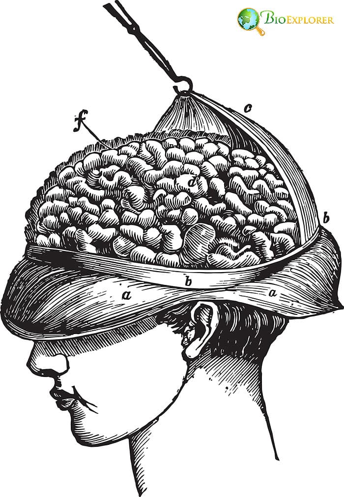 Our brains are incredibly complex. Each area is responsible for specific functions – controlling movements, balance, language, decisions, and overall control. The brain is also highly adaptable and can form new connections and restore necessary networks of one or more of its areas that are damaged or removed due to surgery.
- A team of researchers led by D. Kliemann at the California Institute of Technology has proven that the level of brain plasticity is even higher than previously thought.
- The researchers have gathered data from six adult patients that have undergone the removal of a cerebral hemisphere (hemispherectomy) in childhood and then compared those data to those of the healthy controls.
- The team was looking at the networks within the brain responsible for:
- vision
- movement
- attention
- emotional/fear responses
- Control networks
- It was discovered that the patients with a removed hemisphere still had functional, working networks required for all the mentioned above processes.
- The scientists have also found more reliable connections between the networks in these patients’ brains.
This study has proven that even with such drastic changes in brain anatomy as removing a whole hemisphere, the internal brain networks still support many complex mental processes. It is especially likely if the surgery was done in early childhood.
![]()
A small, well-nourished brain: a new method for producing brain organoids with blood vessels proposed
 The complexity of the human brain makes it very hard to study. Recently, with the emergence of new cell engineering technologies, it has become possible to grow “mini-brains” – so-called brain organoids. Until recently, creating a brain organoid with blood vessels was impossible.
- A team from the Yale Stem Cell Center of the Yale School of Medicine led by Park In-Hyun has proposed a new method of development of brain organoids.
- For this experiment, the researchers have engineered the stem cells to produce a special factor called human ETS variant 2 (ETV2).
- The engineered stem cells were then used to develop human brain organoids in culture.
- Due to the presence of ETV2, microscopic blood vessels were formed inside the organoids.
- Moreover, several blood-brain-barrier properties were observed in the newly formed organoids.
- The blood-brain barrier is a fundamental structure in the brain that protects it from infections without jeopardizing brain nutrition. No previously developed brain organoids had displayed those features.
The unique method proposed by this team of scientists would allow studies of infections that overcome the blood-brain barrier and would also help understand how the blood supply of the brain works.
![]()
These cells are a pain: a new type of cells responsible for pain perception found
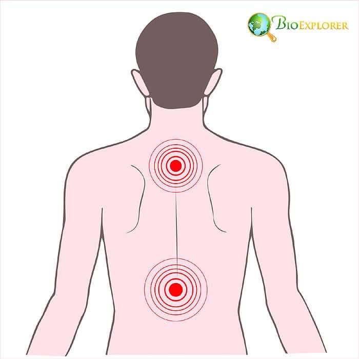 Until recently, it was thought we have multiple nerve endings in the skin that transmit pain signals when our skin is injured, or we get a burn. Abdo et al. have shown that this concept was wrong.
- A team of researchers at Karolinska Institutet, Sweden, led by Hind Abdo, has discovered a novel structure in the skin that is specialized for sensing and transmitting information about pain.
- The structure is composed of a glial cell type with multiple projections.
- The cell projections form a mesh under the skin.
- These cells are also connected to the sensory nerve endings in the skin. When they get stimulated, they use those connections to transmit pain information.
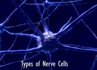
Explore Different Types of Nerve Cells
This discovery completely overturns the previous concepts about pain transmission from the skin.
![]()
A new stage in 3D printing: It is now possible to make a skin graft that has blood vessels with the help of 3D bioprinting
 Three-dimensional printing has stepped into a new era. It is now possible to use bio-inks that contain viable cells instead of plastic or another medium. This approach can potentially be used to print organs for transplantation and grafting.
- The team at the Rensselaer Polytechnic Insitute led by Tania Baltazar has decided to focus on skin.
- Skin grafts are crucial in many cases – for example, for burn victims or people with non-healing skin ulcers.
- Currently, skin transplants may not graft appropriately because they lack blood vessels that could integrate with the patient’s body.
- The team has printed the new type of graft using several cell types:
- fibroblasts and keratinocytes that form the actual skin,
- endothelial cells and placental pericytes that interact together and form blood vessels.
- This approach has resulted in a multi-layered skin graft that had all the major skin components: epidermis, dermis, and functional blood vessels.
Though printing a skin graft with functional blood vessels is a breakthrough, there is still a hurdle before such grafts can be applied. To avoid the possibility of transplant rejection, the cells used in printing need to be engineered with CRISPR help so the resulting graft would suit a particular patient.
![]()
Disappearing muscles: a new study has discovered a new set of muscles in developing human embryos that are similar to those in lizards
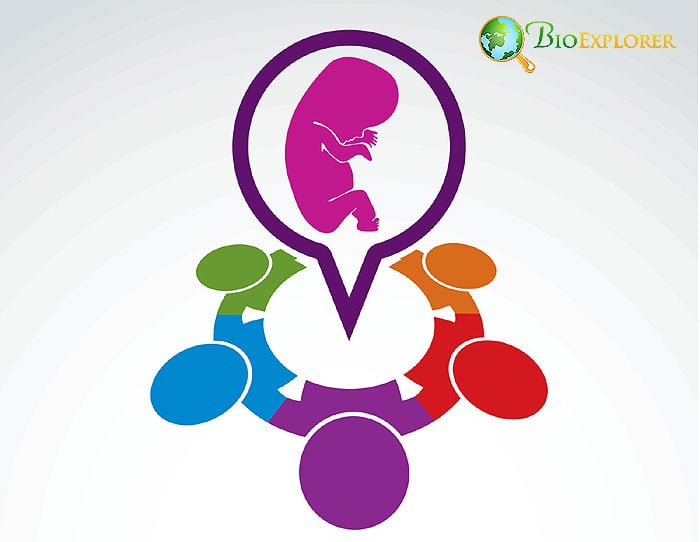 One of the arguments used by Darwin to prove his evolutionary theory was the presence of atavisms: vestigial limbs in snakes, vestigial wings in ostriches, or the birth of human babies covered with hair. Specific organs, like the notochord, appear during the early embryonic stages of animal development and disappear later. Most of those structures were observed in animal embryos but not in human ones.
- Ryogo et al. have shown the presence of atavistic muscles during embryonic development for the first time in humans.
- With the use of new imaging techniques, his team has discovered a new group of muscles in the limbs of developing embryos, called dorsometacarpales.
- These muscles can only be seen in lizards or humans with developmental defects and are absent in healthy adults.
- About 30 muscles of this type present in both hands and feet were registered on the scans of the embryos at about 7 weeks of gestation.
- With time, these muscles are fused or completely disappear by the 13 weeks of gestation.
This is the first time these muscles were found during embryonic development. This discovery may further the specialist’s understanding of congenital muscle problems seen at the later development stages and birth. It is also another piece of proof that evolution is indeed real.
![]()
A secret capillary: a new type of blood vessel discovered in our long bones
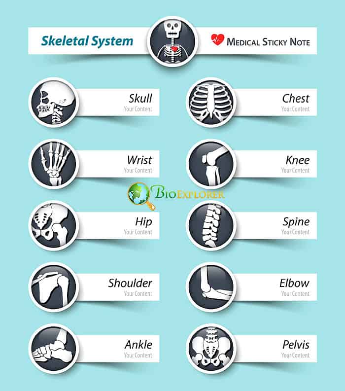 The bones both require blood supply and contribute to the turnaround of red and white blood cells. Previously, it was thought that bones have only a few blood vessels responsible for the transport of cells and nutrients.
- A study performed on the femurs (hip bones) of mice undertaken by a team of researchers at the University Hospital in Erlangen, Germany, has led to a new network of blood vessels.
- With the help of field emission microscopy, the researchers have found a new type of capillary-like vessels that cross the entire long bone perpendicularly that they have named transcortical blood vessels (TBV).
- The TBV network contains both veins and arteries.
- This network plays a crucial role in transporting immune cells from the bone marrow.
- This discovery completely upends the previous concept of anatomy. It provides scientists with new information about the transport of immune cells from the bone marrow to other body areas.
![]()
A new approach to analyzing ultrasound data allows seeing pores inside the bones
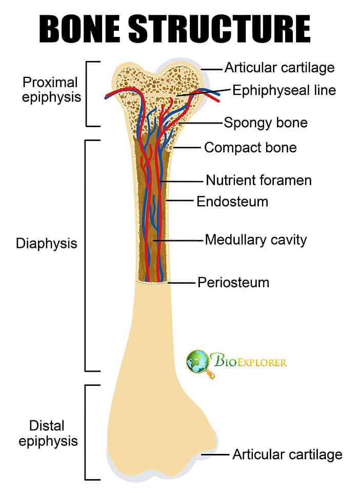 Ultrasound is not used for studying bone because the signal does not go through the solid tissues. The Faculty of Mechanical and Aerospace Engineering at North Carolina University team thought that this might not be a disadvantage in some instances.
- The researchers have found that by analyzing how the signal from the ultrasound scatters in the bone, it is possible to find if there are spaces in the bone itself.
- The presence of pores can be a symptom of an illness – osteoporosis.
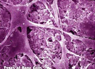
Types of Bone Cells
If this new approach is used at clinics, patients can evade unnecessary X-rays and still get information about whether they have osteoporosis and if their treatment is effective. This is also a good and safe way to study bone structures.
![]()
An underestimated organ: it was proven that the clitoris plays a vital role in the reproduction process
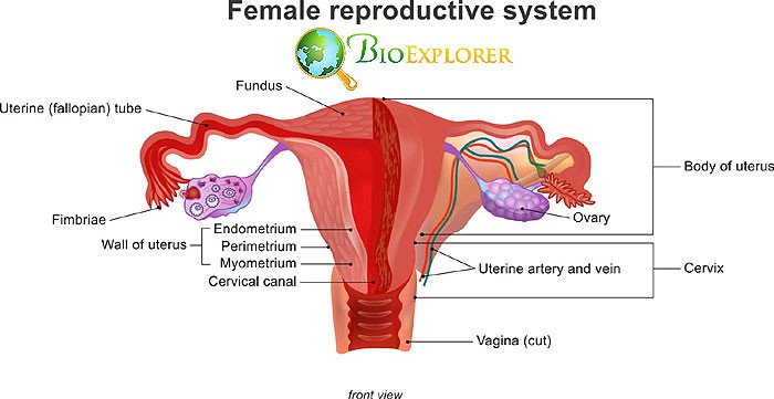 The clitoris is one of the most controversial organs in the female body. Many scientists have time and again postulated that this organ exists simply for initiating sexual pleasure. Several studies performed in the course of the 20th and 21st centuries prove otherwise.
- An independent investigator from Sheffield, United Britain, Roy J.Levin, has systematized the studies devoted to the function of the clitoris that was published over two centuries.
- He has found proof that stimulation of the clitoris during the sexual act initiates a chain of essential processes:
- An increase in the blood flow to the vagina
- Vaginal lubrication
- changes in pH in the vagina
- synthesis of factors that promote sperm activation.
- The researcher points out that the clitoris is crucial for successful reproduction, as its stimulation leads to the overall preparedness for fertilization.
Top 25 Reproductive System Fun Facts
Most of these findings were done several decades prior. However, this review is the most substantiated attempt to prove this point using overwhelming evidence.
![]()
Our future changes our bones: fabella is more prevalent in the population of the 21st century compared to the previous centuries
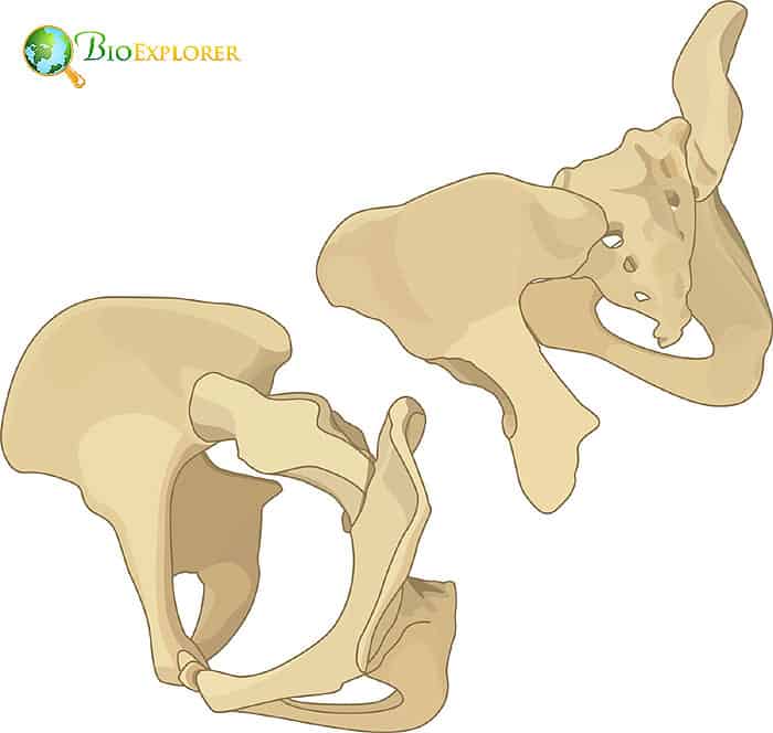 Fabella is a small, sesamoid bone located behind the lateral projection of our femoral bones. It has been known to scientists since 1875.According to observations, fabella is present more often in Asians than non-Asians.
- The systemic analysis of the scientific literature that spans the period from 1875 to 2018 undertaken by Michael A. Bertaume et al. has shown that the fabella occurs significantly more in men and women of the 21st century than in people of previous periods.
- The scientists think this phenomenon is linked to multiple factors.
- One of the reasons is that in the 21st century, people are far more likely to grow bigger and heavier because of access to quality nutrition, which may require additional bones in the knee joint for support.
- The appearance rates of other known sesamoid bones were not altered in the modern population.
This information may be crucial for doctors as the presence of the fabella is associated with knee pain and osteoarthritis.
![]()
Focus on the cranium: new 3D models successfully teach anatomy students about skull development

- Technologies change the way anatomy students learn their subjects. Previously, the students needed to rely on old bone and organ samples or used 2D anatomical atlases, which did not reflect most anatomical structures realistically. Current progress in 3D graphics adds new options for the old subject.
- A new approach to teaching students about skull growth and development was tested During the collaboration between Taibah University in Saudi Arabia and Glasgow University in Glasgow, Scotland, United Kingdom.
- Specimens from Glasgow’s Museum of Anatomy were scanned and digitized.
- Based on the scanned specimens, full-color 3D models of the stages of human skull development were created.
- The models were used as a learning tool during lectures devoted to developing the juvenile skull.
- A student survey was conducted afterward, and the new application was reviewed positively.
The new tool has helped students understand the skull’s development better, and most students liked the idea more than using 2D pictures or specimens in class. Moreover, the approach helped preserve fragile anatomical specimens stored for teaching purposes.
![]()
Changing the way to learn: a student-oriented approach called the jigsaw method was successfully used for teaching complex anatomical subjects to students
- Approach to teaching anatomy is very much the same as it used to be in previous centuries: the students have to learn the structures by heart. They can get additional information from specimens, dissections, and anatomical atlases. However, this type of learning is passive and does not promote full retention of information or critical thinking. A new teaching method based on another active learning approach was tested in India.
- The professors have chosen an active learning concept called jigsaw method for teaching anatomy-related subjects to students.
- The jigsaw method involves separating the subject into several subtopics or assignments and letting the students rotate between subtopics in small groups.
- In the study, two topics were chosen for the new learning approach: the anatomy of the stomach and the anatomy of the uterus.
- Before the test, the students had already had two traditional lectures on the topic and were instructed in the features of the jigsaw method with the school faculty.
- The jigsaw technique, in this case, was organized as follows:
- Both topics were broken down into 5 subtopics.
- The students were first organized into so-called “home groups“, 5 people in each group.
- Each member of the group was randomly assigned to one “expert group.”
- Each expert group was assigned a subtopic, which all the students researched with the help of available sources: Internet search, textbooks, atlases, and other tools.
- After completing each assignment, the students returned to their home groups. They taught each other the information that they had gained previously.
- The feedback from the students and the faculty resulted in a positive evaluation of this teaching approach.
Implementing the jigsaw method was successful but was also deemed to need better organization as it was time-consuming. Also, students applied themselves differently to the assignments, and some shirked their responsibilities. Overall, the method promotes critical thinking and makes the students participate actively in getting and understanding information.
![]()
The smell is still there: several women were found to lack olfactory bulbs but still able to perceive smells
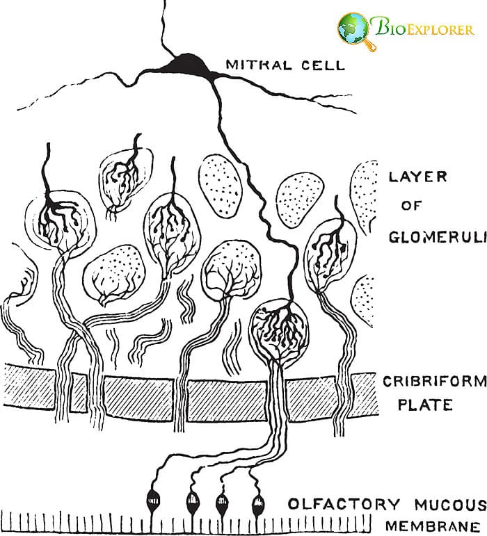 Our brains have specific “first responder” sites that are the first to obtain signals from our sensory organs. Such a site for obtaining smell information is called an olfactory bulb located in the forebrain. A team of scientists at the Weizmann Institute, Rehovot, Israel, led by Noam Sobel, has discovered that olfactory bulbs may sometimes be replaceable.
- During routine MRI studies, a team of Israeli researchers has found two healthy women, who were also left-handed, that did not have olfactory bulbs.
- During the follow-up to this discovery, the team established that despite the lack of those structures, both women could perceive smells during an odor test.
- The data analysis obtained during the Human Connectome Project revealed three more women who lacked the olfactory bulbs and could perform an odor test.
- The researchers proposed that the lack of olfactory bulbs may be present in approximately 0.6% of women in the general population.
- They also propose that the lack of visible olfactory bulbs may be related to left-handedness.
This study proves humans’ high level of brain plasticity. It is also possible that the olfactory bulbs were not seen in MRIs because they were underdeveloped.
![]()
It is not too late to revive a brain: scientists partially revive pig’s brain 4 hours after slaughter using a novel system [USA, April 2019]
Doctors usually proclaim the patient dead when their brain activity ceases. A group of researchers who undertook a recent experiment at Yale University shows that stating death can be more complicated than that.
- The researchers have developed a novel perfusion system that works with organs outside the body.
- They have also produced a blood substitute with several useful properties:
- The substitute was based on hemoglobin.
- The substitute did not contain any cells.
- The substitute could restore oxygen supply to the brain and protect brain cells.
- The product also could prevent brain swelling.
- Brains taken from pigs slaughtered 4 hours prior were used in the experiment.
- The brains were connected to the new system, and the scientists observed the following changes:
- Most of the brain cells remained intact.
- Cell death in the brain has declined.
- The nerve and brain blood vessel cell responses were restored.
The brain has a previously undiscovered potential for restoration, which may be used, for example, in brain transplant surgeries of the future.
![]()
Suggested Reading:
Top 10 Anatomy/Physiology News in 2020
Many discoveries made in the field of anatomy were due to the progress in new technologies and approaches. The specialists currently have a range of modern imaging and biochemical techniques that allow us to see the anatomical structures significantly better.
These new methods also allow us to look with a less biased view into the past – and evaluate the contribution of the previous generations more objectively, as was the case of Lorenzo Tencini.
Without a proper understanding of basic anatomy, training doctors or developing therapies is impossible. That is why anatomy remains a highly relevant science even after many centuries of studies in this area.
![]()


