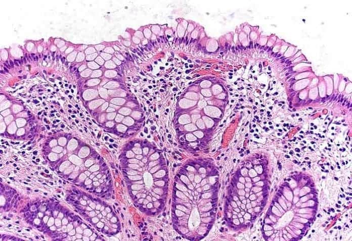
These cells, mostly composed of secretory cells employ a so-called “mucosal barrier ” that serves as a lubricant and aids in the preservation of the epithelium. In particular, cells known as goblet cells are an important component in this barrier and constitute the majority of the immune system
In the last article, we looked at the Kupffer Cells
How to pronounce Goblet Cells? Goblet Cells 🔊 Goblet Cells Word Origin: Celtic, Middle-English
What are Goblet Cells? Goblet Cell Anatomy (Source: Wikimedia) goblet-like shape [1] after they collapse following mucin secretion.[2] are dependent on their age. Young cells are rounded but increase in size and flatten as they age.
Goblets cells have a very prominent morphology; having the nucleus, mitochondria, Golgi body, and the endoplasmic reticulum at the basal portion of the cell. The rest of the cell is filled with mucus in secretory granules. When fixed, these cells appear to have a narrow base and expanded apical portion that extends up to the lumen.
Where are Goblet Cells Found? Goblet Cells Location (Source: Wikimedia) [3] of many organs. In particular they are found lining the epithelium of respiratory organs (trachea, bronchioles, and bronchi), digestive organs (small and large intestines), and the conjunctiva in the upper eyelid. Among all of these organs, they are most abundant in the intestines.
Origin & Development of Goble Cells Goblet cells along with other principal cells in the gastrointestinal tract, (i.e. enteroendocrine cells, enterocytes, and Paneth cells), emerge from the multipotent cells (PDF ) (cells that can give rise to different cell types) in the base of the Crypts of Lieberkühn.
In humans, these in general appear during the fetal development of the small intestine at the 9th to 10th weeks gestation. The overall morphology of these cells is created by the distended theca, the sheath of cells that covers the structure, that contains mucin granules found below the apical membrane. Functions of Goblet Cells Apart from comprising the epithelial lining of various organs, production of large glycoproteins and carbohydrates, the most important function of goblet cells is mucus secretion. This mucus is a gel-like substance that is composed mainly of mucins, glycoproteins, and carbohydrates.
The following are the functions of mucus.
1. Secretion in the small and large intestine According to a study published in the
American Journal of Physiology [4] they found in the small and large intestine are able to synthesize and maintain the so-called mucus blanket that in turn produces glycoproteins known as mucins.
These mucins help neutralize the acids produced by the stomach. They also help in lubricating the epithelium for the easier passage of food. Although the production of mucus is the main function of them, a recent study published in the journal Mucosal Immunology [5] have shown that goblet cells in the small intestine can accumulate and uptake antigens (toxins that induces an immune response). In the large intestine, the formed mucus blanket/barrier [6] inhibits inflammation by preventing the passage of luminal bacteria and food-derived antigens from passing through it. Such phenomenon is called as the oral tolerance.
2. Secretion in the respiratory tract While most cells in the respiratory tract are ciliated columnar cells, there are some goblet cells present in the epithelia. In these locations, they are situated with their apices protruding into the lumen in order to react rapidly whenever a chronic airway insult happens or a foreign body is inhaled.
A study published in the journal European Respiration Journal [7] revealed that the mucins produced by the goblet cells are responsible for the trapping and transport of the inhaled foreign bodies (i.e. allergens, particles, and microorganisms). Aside from that, this study also showed that goblet cells can even produce more mucus than any other glands in the body.
3. Secretion in the conjunctiva In the eyes, the conjunctiva is the thin semi-transparent membrane that covers the exposed areas of the sclera (eyeballs) and the inner surface of the eyelids.
Organs, like the conjunctiva, that come in contact with the exterior environment are lined with goblet cells that function for lubrication (along with the secretion of tears) against foreign debris and agents.
How Do They Secrete Mucus? As mentioned earlier, goblet cells secrete mucus through merocrine secretion, which in turn serves a variety of functions. But in the first place, how do these cells secrete such powerful substance?
The secretion of mucus [3] is preceded by a stimuli. Along with the secretory granules, they secrete the mucus via exocytosis (process where the contents of the vacuole is released).
When inside the goblet cell, the mucus is initially in a condensed state. However when it gets released, it dramatically and instantaneously expands. This is apparently true because according to studies, the mucin gel in goblet cells can expand up to 500 times its original volume in just 20 milliseconds!
Diseases Associated With Goblet Cells Because they can serve as progenitor (can give rise to) other cells, they can also be a good indicator of the condition of their neighboring cells, tissues, or organs.
According to some studies [8] , goblet cells are associated with diseases in the respiratory tract like cystic fibrosis and chronic bronchitis. In these type of diseases, they can either undergo metaplasia ( change to other type of cells) or hyperplasia (abnormal increase of number).
While goblet cells can perform functions that are beneficial to an organism, too much proliferation of it is not good either. When both goblet cells and neuroendocrine cells are produced, tumors called goblet cell carcinoids may arise.
This tumor occurs in the appendix and may even require removing the said organ when the situation gets worse.
In general, goblet cells and the mucus they produce have long been poorly studied and appreciated. Many other important questions about these cells remain.
Hence more detailed studies
Cite This Page
[1] – goblet cell. Dictionary.com. Dictionary.com Unabridged. Random House, Inc. Link (accessed: December 15, 2016). [2] – “the laboratory fish ” by Gary K Ostrander, John Hopkins University, MD – Book link . [3] – “What is the function of goblet cells? ” What is the function of goblet cells? Accessed December 15, 2016. Link . [4] – “Functional biology of intestinal goblet cells. ” The American journal of physiology. Accessed December 15, 2016. Link . [5] – Nature.com. Accessed December 15, 2016. Link . [6] – Shan, Meimei, Maurizio Gentile, John R. Yeiser, A. Cooper Walland, Victor U. Bornstein, Kang Chen, Bing He, Linda Cassis, Anna Bigas, Montserrat Cols, Laura Comerma, Bihui Huang, J. Magarian Blander, Huabao Xiong, Lloyd Mayer, Cecilia Berin, Leonard H. Augenlicht, Anna Velcich, and Andrea Cerutti. “Mucus Enhances Gut Homeostasis and Oral Tolerance by Delivering Immunoregulatory Signals.” Science (New York, N.Y.). 2013. Accessed December 15, 2016. Link . [7] – “Airway goblet cells: responsive and adaptable front-line defenders. ” The European respiratory journal. Accessed December 15, 2016. Link . [8] – “Goblet Cells. ” Goblet Cells. Accessed December 15, 2016. Link . 
