What is Meiosis?
Jump to:
As a general rule, all cells come from pre-existing cells and the primary mode to how this happens is through the process of cell division. In this process, the parent cell divides into daughter cells while at the same time passing its genetic material across generations.
In eukaryotic organisms, two kinds of cell division exist: mitosis and meiosis. On one hand, mitosis is concerned in the production of daughter cells that are mere copies of each other. On the other hand, meiosis involved the production of the reproductive cells that bear unique genetic characteristics.
At the level of the genes, sexual reproduction is focused on the fusion of the paternal and maternal genes that result in a new combination of genes. And this is where the process of meiosis becomes important.
In this article, discover how meiosis occurs in living cells, its different stages, and its significance in the survival of eukaryotic organisms.
What is Meiosis?
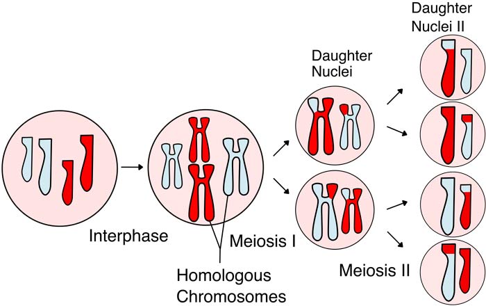
Meiosis[1] is a type of cell division that involves the reduction in the number of the parental chromosome by half and consequently the production of four haploid daughter cells. This process is very essential in the formation of the sperm and egg cells necessary for sexual reproduction. When the haploid sperm and egg fuse, the resulting offspring acquires the restored number of chromosomes.
-
Meiosis is highly ubiquitous among eukaryotes as it can occur in single-celled organisms like yeast as well as multi-cellular ones like humans.
-
The process of meiosis is very essential in ensuring genetic diversity[2] through sexual reproduction.
-
In humans, two distinct types of daughter cells are produced by males and females (sperm and egg cells respectively).
![]()
Stages of Meiosis
Contrary to mitosis, meiosis[3] involves the division of diploid parental cells (both paternal and maternal) into haploid offspring with only one member of the pair of homologous chromosome from the parents.
1. Meiosis I
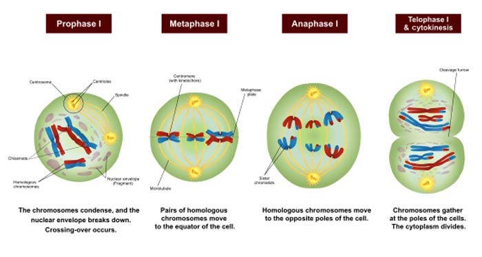
Just like in mitosis, a cell must first undergo through the interphase before proceeding to meiosis proper. It increases in size during G1 phase, replicates all the chromosomes during S phase, and makes all the preparations during the G2 phase. Like the usual mitosis, the first meiotic division is divided into four different stages: prophase I, metaphase I, anaphase I, and telophase I.
Prophase I.
By far, prophase[4] I of meiosis is considered as the most complicated step in the whole process. As compared to mitotic prophase, the prophase of meiosis is definitely longer. In this stage of meiosis I, the chromosomes start to condense and pair up with its homologue. Basically, the first meiosis begins with a very long prophase that is divided into five phases: leptotene, zygotene, pachytene, diplotene, and diakinesis.
Leptotene
Leptotene is the first stage of prophase during meiosis I. This phase is characterized by the condensation of the chromosomes wherein they become visible as chromatin. It comes from the two Greek words “lepto” and “tene” which mean “thin” and “ribbon” respectively.
Zygotene
Coming from the Greek words “zygo” and “tene” which mean “union” and “thread“, zygotene is the second phase of prophase I. During this stage, homologous chromosomes begin to form an association called a synapse which results to pairs of chromosomes that has four chromatids.
Pachytene
This is the phase where the crossing over between pairs of homologous chromosomes occurs. The structure formed is referred to as the chiasmata. In contrast with leptotene (“thin thread“), the Greek word “pachy” means “thick“; thure referring to the characteristic of the chromosome in this stage.
Diplotene
In this phase, the separation of the homologous chromosomes is starting but they remain attached through the chiasmata. The word “diplo” in diplotene means “double“.
Diakinesis
Following diplotene is the final phase called diakinesis, which comes from the Greek words “dia” which means “across” and “kinesis” which means “motion“. This is when the homologous chromosomes continue to separate as the chiasmata move to the opposite ends of the chromosomes.
Metaphase I.
In this stage, the homologous pairs of chromosomes randomly align at the metaphase plate.[5] Such configuration becomes the source of genetic material as the chromosomes from the male and female parents appear similar but are not exactly identical.
Anaphase I.
The homologous chromosomes become separated as they are pulled toward the opposite ends[6] of the cell. However, the sister chromatids remain attached to their pair and do not move apart.
Telophase I.
Like in the telophase of mitosis, the chromosomes finally are separated at the different sides of the cell. In addition, the chromosomes return to their uncondensed forms as the nuclear membrane is reformed. The division of the cytoplasm (referred to as cytokinesis) occurs simultaneously with telophase I, resulting to two haploid daughter cells.
![]()
2.Meiosis II
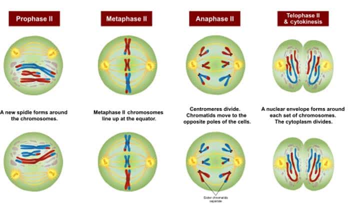
Cells undergo through meiosis I to meiosis II without the replication of the genetic material. It is important to note that the cells that undergo meiosis II are the daughter cells produced during meiosis I. Meiosis II is shorter than meiosis I but still is divided into four stages: prophase II, metaphase II, anaphase II, and telophase II.
Prophase II.
Prophase II is almost similar to mitotic prophase. In prophase II, the nuclear envelope disintegrates as the chromosomes condense[7]. Aside from that, the centrosomes separate with each other while the spindle fibers try to catch the chromosomes.
Metaphase II.
Just like in meiosis I, meiosis II[8] is when the chromosomes align at the metaphase plate because of the attachment of the spindle fibers to the centromeres of chromosomes. The segregation of different types of chromosome is what creates the difference between the two metaphases of meiosis.
Anaphase II.
Unlike anaphase I, which involves the separation of homologous chromosomes, anaphase II is the separation of sister chromatids. In this stage, chromosomes (each with a chromatid) are separated from each other as they move toward the opposite poles of the cell.
Telophase II.
Just like the usual telophase, the cell cytoplasm divides equally and shortly after is reformed. The result of telophase II is the formation of four haploid cells (having each set of chromosomes). However, it is important to note that the four daughter cells are not identical with each other due to the events in meiosis I (i.e. random crossing over and random alignment of chromosomes.
![]()
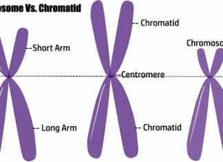
What is the Difference Between Chromosome and Chromatid?
Errors In Meiosis: The Science Behind Nondisjunction
![]()
For additional illustrations, you may want to check out this YouTube video on meiosis:
Importance of Meiosis
-
One of the most important purposes of meiosis[9] is the generation of genetic diversity among individuals. This fact becomes very crucial as such diversity allows the development of certain adaptations that will help in surviving in an ever-changing environment. Such recombination is essential in the repair and regulation of genetic abnormalities that tend to arise in sex cells.
-
Another importance of meiosis is that it helps in the reprogramming of the sex cells, giving rise to the zygote. Lastly, meiosis is essential for the maintenance of the immortality of the sex cell line which is possible due to the removal and rejuvenation of defective RNA and proteins.
![]()
But come to think about it. Isn’t it fascinating how a single cell is able to carry out a very accurate and highly conserved physiological process?
Cite this page
Bio Explorer. (2026, January 3). What is Meiosis?. https://www.bioexplorer.net/divisions_of_biology/cell_biology/meiosis/
References
- [1] – “meiosis | Learn Science at Scitable”. Accessed October 01, 2017. http://www.nature.com/scitable/definition/meiosis-88.
- [2] – “What Is Meiosis?”. Accessed October 01, 2017. http://www.livescience.com/52489-meiosis.html.
- [3] – “Meiosis – Molecular Biology of the Cell – NCBI Bookshelf”. Accessed October 01, 2017. https://www.ncbi.nlm.nih.gov/books/NBK26840/.
- [4] – “Substages of Prophase I – Online Biology Dictionary”. Accessed October 01, 2017. http://www.macroevolution.net/prophase-details.html.
- [6] – “Meiosis | Cell division | Biology (article) | Khan Academy”. Accessed October 01, 2017. https://www.khanacademy.org/science/biology/cellular-molecular-biology/meiosis/a/phases-of-meiosis.
- [7] – “Life Sciences Cyberbridge”. Accessed October 01, 2017. http://cyberbridge.mcb.harvard.edu/mitosis_7.html.
- [9] – “The biological significance of meiosis. – PubMed – NCBI”. Accessed October 01, 2017. https://www.ncbi.nlm.nih.gov/pubmed/6400219.

This article was very awesome…..